ARTÍCULOS ORIGINALES
The influence of maxillary and mandibular osteoporosis on maximal bite force and thickness of masticatory muscles
Paulo B. Vasconcelos1, Marcelo Palinkas2, Luiz G. de Sousa1, Simone C. H. Regalo1, Carla M. Santos1, Moara de Rossi1, Marisa Semprini1, Priscilla H. Scalize1, Selma Siéssere1
1 Department of Morphology, Physiology and Basic Pathology, Ribeirão Preto Dental School,
University of São Paulo, Ribeirão Preto, Brazil
2 Department of Restorative Dentistry, Ribeirão Preto Dental School, University of São Paulo,
Ribeirão Preto, Brazil
CORRESPONDENCE Dr.Marcelo Palinkas Department of Restorative Dentistry Avenida do Cafe, s/n, Ribeirao Preto, Sao Paulo, Brazil e-mail: palinkas@usp.br
ABSTRACT
The aim of this study was to examine the bite force and masseter and temporal muscle thickness in individuals with maxillary and mandibular osteoporosis. 72 individuals were distributed into two equal groups: (1) facial osteoporosis and (2) healthy controls. Bite force on the right and left molar regions was recorded with a dynamometer and the highest value out of three measurements was recorded as the maximal bite force. Muscle thickness was measured with a SonoSite Titan ultrasound scanner. Ultrasound images were obtained of the bilateral masseter and temporal muscles at rest and at maximal voluntary contraction. The means of the measurements in each clinical condition were analyzed with multivariate statistical analysis (SPSS 19.0). Student's t test indicated no significant difference for muscle thickness, but indicated significantly lower bite force values in the osteoporosis group (p>0.05). Lower bite force in individuals with facial bone loss demonstrates functional impact of osteoporosis on the complex physiological stomatognathic system.
Key words: Osteoporosis; Ultrasound; Bite force; Masticatory muscles.
RESUMO
A influência da osteoporose maxilar e mandibular na força de mordida e espessura dos músculos mastigatórios
Este estudo teve como objetivo analisar a forca de mordida e a espessura dos musculos masseter e temporal em individuos com osteoporose maxilar e mandibular. 72 individuos distribuidos em dois grupos equivalentes: (1) osteoporose facial e (2) controles saudaveis. Forca de mordida nas regioes de molar direita e esquerda foi gravada com o dinamometro e o valor mais alto das tres medidas foi registrado como a forca de mordida maxima. A espessura muscular foi mensurada com ultrassom SonoSite Titan. As imagens de ultrassom foram obtidas dos musculos masseter e temporais bilateral em repouso e em contracao voluntaria maxima. As medias das medidas em cada condicao clinica foram analisadas com a analise estatistica multivariada (SPSS 19.0). Teste t de Student nao revelou diferencas significativas para a espessura musculos, mas indicou valores significativamente mais baixos de forca de mordida no grupo com osteoporose (p> 0,05). Forca de mordida menor em individuos com perda ossea facial demonstra um impacto funcional da osteoporose na fisiologia complexa do sistema estomatognatico.
Palavras-chave: Osteoporose; Ultrassom; Forca de mordida; Musculos mastigatorios.
INTRODUCTION
Increasing longevity of the world population has
led to osteoporosis being considered the "epidemic
of the twenty-first century" 1,2. Osteoporosis is a
serious public health problem for middle-aged and
elderly women and increases after menopause 3. By
2050, the worldwide incidence of hip fracture is
projected to increase by 240% for women and 310%
for men. The estimated number of osteoporosis hip
fractures worldwide is expected to rise from 1.66
million in 1990 to 6.26 million in 2050, even if ageadjusted
incidence rates remain stable 4. The International
Osteoporosis Foundation in 2013 reports
that it is one of the most important diseases associated
with aging.
Systemic osteoporosis affects femoral, radial, and
spinal bones, in addition to affecting craniofacial
bones and oral structures, directly influencing vari-
ous oral conditions and dental procedures 5,6. Clinical
and scientific dental interest in the effects of
osteoporosis on facial structures has been growing.
In a preliminary study, Siessere et al. 7 evaluated
the electromyographic activity of the masseter and
temporal muscles of patients with maxillary and
mandibular osteoporosis compared to a control
group. They found that the decrease in the amount
of maxillary and mandibular bone tissue that supports
the muscle structure in individuals with osteoporosis
does not cause a change in the level of
electromyographic pattern activation.
Dental radiographs might be useful for screening
for osteoporosis. Some studies indicate the use of
the relationship between mandibular bone mineral
density (BMD) and other skeletal sites commonly
used for bone densitometry in the detection of
osteoporosis 8. The evaluation of dental radiographs
may have a role in the detection of individuals with
osteoporosis 9. Other oral signs of osteoporosis
could be alveolar ridge resorption, tooth loss and
chronic destructive periodontal disease 10.
Mastication is one of the functions of the stomatognathic
system, which comprises a functional and
physiological entity integrating a set of organs and
tissues whose biology and physiopathology are
absolutely interdependent and therefore require
complex evaluation. In addition to the electrical
activity previously evaluated 7, structural evaluation
of masticatory muscles and their ability is
essential for complete understanding of the possible
influences of osteoporosis on the masticatory
process.
In this context, the aim of this study was to investigate
the thickness of the masseter and temporal muscles
and the bite force of patients with mandibular
and maxillary osteoporosis. The data from these
osteoporotic patients was compared to data obtained
from healthy individuals.
MATERIALS AND METHODS
Volunteers
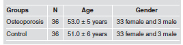
Seventy-two individuals of both genders, with an
average age of 53.0}5 years, with no distinction of
ethnicity or social class, took part in this study.
They were divided into two groups of 36. Group 1
consisted of thirty-six individuals selected at random
from the pool of users of the Radiology Clinic
at the Ribeirao Preto Dental School, University of
Sao Paulo, Brazil, with mandible and maxillary
osteoporosis, who had been diagnosed by means of
panoramic radiographs, obtained through the acquisition
of digital image indirectly, with the chassis
plans 15x30 or 18x24cm, using the panoramic Xray
machine and cephalometric brand - Siemens,
model - Orthophos CD with kVp: 90 mA and: 16
and turnover time 14.1s. Lorente – Ramos et al. in
2011 reported that panoramic radiographs showed
low bone mineral density (BMD), confirmed by
BMD values of the lumbar spine (L1–L4) as measured
by the exam Dual Energy X-ray Absorptiometry
or DEXA, which has high diagnosis accuracy
and a low dose of radiation compared to other methods.
The DEXA exam was used to diagnose skeleton
osteoporosis in each individual. The scanner
takes a picture of the bones in the spine, hip, total
body and wrist, and calculates their density. To take
a DEXA bone density scan, the patient lies on a bed
underneath the scanner, a curving plastic arm that
emits X-rays. These low-dose X-rays form a fan
beam that rotates around the patient. During the test,
the scanner moves to capture images of the patient's
spine, hip or entire body. The test takes about 20
minutes to perform and is painless. Group 2 (control)
included thirty-six individuals, who were
employees, and relatives of patients and students,
paired subject-to-subject by gender and age (Table 1)
with the subjects with osteoporosis.
Table 1: Demographics of the two groups evaluated.
Age, gender and standard deviation (±) in
osteoporosis and control group.
The sample and inclusion/exclusion criteria were selected by means of anamneses and clinical examinations. The anamneses provided information on the participants' personal data, medical and dental history, any existing parafunctional habits, and possible temporomandibular dysfunction symptoms. All subjects were completely dentate or orally rehabilitated by means of partial fixed dentures or dental implants and had no periodontal problems. The following exclusion criteria were applied during the anamnesis: any systemic or local disorders other than osteoporosis, which could compromise craniofacial growth or the masticatory system, such as neurological disorders, cerebral palsy, and others; taking any medication that could interfere with muscle activity, such as antihistamines, sedatives, homeopathy, or central nervous system depressors; being under any kind of treatment that could, directly or indirectly, interfere in muscle activity during the period in which the study was performed, such as speech therapy and otorhinolaryngology treatment. Subjects were informed about the purposes and stages of the study and they all provided written consent, signing the form previously approved by the National Health Council (process number 2006.1.242.58.3). Thirtysix control patients were matched individual to individual with the osteoporosis sample. Each subject was assigned to one of two groups, named 1 and 2, and only one examiner knew which group the numbers referred to (control or osteoporosis). All examinations were performed without the researchers knowing which group the subjects belonged to, which made it a double-blind study.
Ultrasound analysis
Muscle thickness was analyzed with a SonoSite Titan
ultrasound tool using a high-resolution real-time
56mm/ 10 MHz linear-array transducer placed transversally
to the muscle fibers. The middle of masseter
muscle was considered to be located between 1.5 and
2.0 cm above the jaw angle towards the upper eyelid,
and the anterior portion of the temporal muscle
between 1.0 and 1.5 cm to the back and above the
external palpebral commissure. The muscle location
was confirmed by palpation and transducer movement
at the time of image acquisition. The ultrasound program
enables measurements with a precision of 0.1
mm. Three acquisitions were made in each muscle
condition (rest and dental clenching at maximal habitual
optimized imaging). Ultrasound images were
obtained from bilateral temporal and masseter muscles
at rest and maximal voluntary contraction. During
the examination, the participants remained seated,
leaning on the backrest with the head unrestrained.
Measurements were taken at intercuspidation, with an
interval of 2 min between each acquisition for the participants
to rest their muscles after dental clenching.
Bite Force analysis
Bite force measurements were collected with the volunteers
sitting on a comfortable chair (office-like),
with arms extended along the body and hands resting
on their thighs. The records were taken with a digital
dynamometer, model IDDK (Kratos, Cotia, Sao
Paulo, Brazil), with a capacity of 1000 N, adapted to
the mouth. The apparatus has a ‘‘set-zero'' key, which
allows the exact control of the values obtained and
also ‘‘peak'' registers that facilitate the record of the
maximal force during measurements. It has two arms
with plastic disks on each end, on which the force to
be measured is applied. Its high precision charge cell
and electronic circuit to indicate force supply precise
measurements easily viewed on a digital display. The
dynamometer was cleaned with alcohol, and disposable
latex finger cots (Wariper, Sao Paulo, Brazil) were
positioned on the biting arms as a biosafety measure.
The participants were given detailed instructions and
bite tests were performed before the actual recordings
were made in order to ensure the reliability of the procedure.
The volunteers were then asked to bite the
dynamometer three times with maximal force, with a
2-min rest interval between records. Evaluations were
performed at the first molars (left and right). Maximal
bite force was measured in N through the ‘‘peak'' force
record indicated on the screen, for subsequent analysis.
The highest value out of three records was considered
as the individual's maximal bite force.
Method Error
The method error of muscle thickness measurements
was performed on 18 individuals. Recordings
were obtained at two different sessions with a
7-day interval. At each session, an average of three
measurements was considered for each side and
used later to assess the results. The method error
(Se) was calculated using Dahlbergs's formula: Se
= √ Σ d2 / 2n, where "d" is the difference between
the two recordings of the individual and "n" the
number of double recordings. Percentage errors
were calculated using the formula % = (Se/mean)
100%, where "Se" is the result from Dahlberg's formula
and mean corresponds to the mean value of
the total of the initial and second measurements. A
small difference was found between the first and
second (1 week later) series (2.57 – 6.37 %).
The method error of bite force measurements was
performed on five subjects. Recordings were
obtained at two different sessions with a 7-day
interval. At each session, the mean of three bites
was considered for each side and used later to assess
the results. Paired measurements were analyzed to
identify systematic errors. No difference was found
between the first and second (one week later) series.
Data analysis and statistics
The maximal molar bite force and muscle thickness
measurements on both sides were analyzed using
Student's T - test (SPSS 19.0 for Windows; Chicago,
USA). A 5% (p≤0.05) level of significance was
adopted.
RESULTS
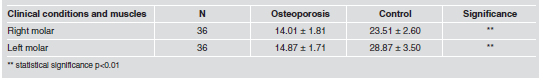
There was no significant difference between the osteoporosis and control groups regarding masseter and anterior temporalis muscle thickness during rest or dental clenching (Table 2). The bite force of the osteoporosis group was statistically significantly lower (p<0.01) than the bite force of the control group (Table 3).
Table 2: Mean, standard deviation (±) and statistical significance of US thickness (mm) of the right and left
masseters (RM and LM) and anterior temporalis (RT and LT) muscles during rest and dental clenching, in osteoporosis and control group.

Table 3: Mean, standard deviation (±) and statistical significance of maximal bite force (N) in osteoporosis
and control group.
DISCUSSION
Ultrasound scanning imaging (US) allows realtime
evaluation of human masticatory muscle morphology.
It is a considerable improvement over
computed tomography and magnetic resonance
imaging because it does not produce cumulative
biological effects, and it has greater clinical availability
and lower cost, making it suitable for largescale
studies 11,12.
The thickness of the masseter and temporal muscles,
as measured by US, has been related to occlusion,
temporomandibular dysfunction, and gender 13.
Thus, this measurement deserves special attention
when studying mastication 14,15. The generalized
bone loss in the skeleton found in osteoporotic
patients can cause disturbances in the masticatory
system, such as modification of muscular position
and masticatory muscle hyperactivity and thus,
increases the chances of temporomandibular or muscular
disorders 7.
It is therefore essential to examine the thickness and
bite force of osteoporotic patients in order to analyze
possible functional changes associated with
this disease.
In the present study, both the osteoporosis and control
groups presented higher masseter and temporalis
thickness during contraction than at rest, which
is in accordance with other studies 11,15-18.
The absence of differences in muscular thickness
between groups also indicates that facial osteoporosis
does not interfere in masseter and temporalis
morphology. According to Siessere et al.7, the masticatory
efficiency of osteoporotic patients is similar
to that of healthy individuals when evaluated by
electromyography. The normal activity of masticatory
muscles may explain the normal thickness of
these muscles.
On the other hand, osteoporosis has a strong association
with the progressive reduction in muscle
mass, strength and function (sarcopenia) 19-22 that
affects older people 23. In age-related muscle atrophy,
a decrease in both muscle fiber size and number
has been reported 24.
The osteoporosis group had significantly lower bite
force than the control group. Because of the reduction
of bone mass, it is suspected that the patients
with osteoporosis tend to have less masticatory
muscle strength than healthy patients. If the musculature
is not trained over several years, there is a
reduction in bite force 25. In one study, a Brazilian
urbanized population was found to have lower bite
force when compared to a Brazilian indigenous
population, because the soft food consumed by the
white population fostered non-trained masticatory
musculature 26. Thus, if osteoporotic patients do not
exert masticatory muscles for a long period, this
reduced function is expected to affect muscular
thickness.
Osteoporosis is a disease that occurs principally in
elderly people. Good nutrition is crucial to the
reduced morbidity of osteoporotic patients. There is
an effective participation of bite force in mastication.
Thus, if bite force increases, masticatory efficiency
increases as well 12,17. Bone tissue is continuously
remodeling in response to mechanical stress. The
alveolar bone mass and the cross-sectional dimension
of the alveolar bone increase with increasing
functional loading 26,27.
A thicker masseter muscle is associated with a higher
local bone density 26. Thus, the maintenance of a
higher muscular loading may contribute to bone
loss control in osteoporotic patients. However, further
studies are required to evaluate the possible
positive effects of muscular stimulation therapy on
the jaw muscles of osteoporotic patients.
This study verified lower bite force in patients with
osteoporosis than in healthy controls. In addition,
both the osteoporosis and control groups presented
higher masseter and temporalis thickness during
contraction than at rest. If bite force is positively
correlated to masticatory efficiency, then it very
important to plan for the treatment of patients with
osteoporosis via the training of masticatory muscle
force as a way to improve masticatory efficiency.
ACKNOWLEDGEMENTS
This study was supported by FAPESP (Grant n° 2006/53563-9).
1. NIH Consensus Development Panel on Osteoporosis Prevention, Diagnosis, and Therapy. Osteoporosis prevention, diagnosis, and therapy. JAMA 2011;14:785-795.
2. Reginster JY, Burlet N. Osteoporosis: A still increasing prevalence. Bone 2006;38:S4-S9.
3. Nanninga GL1, de Leur K, Panneman MJ, van der Elst M, Hartholt KA. Increasing rates of pelvic fractures among older adults: The Netherlands, 1986-2011. 2014, DOI: 10.1093/ageing/aft212.
4. Benson BW, Prihoda TJ, Glass BJ. Variations in adult cortical bone mass as measured by a panoramic mandibular index. Oral Surg Oral Med Oral Pathol 1991;71:349-356.
5. Dervis E. Oral implications of osteoporosis. Oral Surg Oral Med Oral Pathol Oral Radiol Endod 2005;100:349-356.
6. Aguirre JI, Akhter MP, Kimmel DB, Pingel J, Xia X, Williams A, Jorgensen M, Edmonds K, et al. Enhanced alveolar bone loss in a model of non-invasive periodontitis in rice rats. Oral Diseases 2012;18:459-468.
7. Siessere S, Lima NdeA, Semprini M, de Sousa LG, Paulo Mardegan IJ, SAC, Regalo SCH. Masticatory process in individuals with maxillary and mandibular osteoporosis: electromyographic analysis. Osteoporos Int 2009;20:1847-1851.
8. Siminoski K, O'Keeffe M, Brown JP, Burrell S, Coupland D, Dumont M, Ganguli SN, Hanley DA, et al. Canadian Association of Radiologists Technical Standards for Bone Mineral Densitometry Reporting. Can Assoc Radiol J 2013; 64:281-294.
9. Mohajery M, Brooks SL. Oral Radiographs in the detection of early signs of osteoporosis. Oral Surg Oral Med Oral Pathol 1992;73:112-117.
10. Esfahanian V, Shamami MS, Shamami MS. Relationship between osteoporosis and periodontal disease: review of the literature. J Dent (Tehran) 2012;9:256-264.
11. Bertram S, Brandlmaier I, Rudisch A, Bodner G, Emshoff R. Cross-sectional characteristics of the masseter muscle: an ultrasonographic study. Int J Oral Maxillofac Surg 2003; 32:64-68.
12. Raadsheer MC, Van Eijden TM, Van Ginkel FC, Prahl- Andersen B. Human jaw muscle strength and size in relation to limb muscle strength and size. Eur J Oral Sci 2004;112:398-405.
13. Palinkas M, Nassar MS, Cecilio FA, Siessere S, Semprini M, Machado-de-Sousa JP, Hallak JE, Regalo SC. Age and gender influence on maximal bite force and masticatory muscles thickness. Arch Oral Biol 2010;55:797-802.
14. Prabhu NT, Munshi AK. Measurement of masseter and temporalis muscle thickness using ultrasonographic technique. J Clin Pediatr Dent 1994;19:41-44.
15. Raadsheer MC, Kiliaridis S, Van Eijden TM, Van Ginkel FC, Prahl-Andersen B. Masseter muscle thickness in growing individuals and its relation to facial morphology. Arch Oral Biol 1996;41:323-332.
16. Kiliaridis S, Kalebo P. Masseter muscle thickness measured by ultrasonography and its relation to facial morphology. J Dent Res 1991;70:1262-1265.
17. Bakke M, Tuxen A, Vilmann P, Jensen BR, Vilmann A, Toft M.. Ultrasound image of human masseter muscle related to bite force, electromyography, facial morphology, and occlusal factors. Scand J Dent Res 1992;100:164-171.
18. Kubota M, Nakano H, Sanjo I, Satoh K, Sanjo T, Kamegai T, Ishikawa F. Maxillofacial morphology and masseter muscle thickness in adults. Eur J Orthod 1998;20:535-542.
19. Walsh MC, Hunter GR, Livingstone MB. Sarcopenia in premenopausal and postmenopausal women with osteopenia, osteoporosis and normal bone mineral density. Osteoporos Int 2006;17:61-67.
20. Scott D, Blizzard L, Fell J. A prospective study of selfreported pain, radiographic osteoarthritis, sarcopenia progression and falls risk in community-dwelling older adults. Arthritis Care Res (Hoboken) 2012;64:30-37.
21. Gould H, Brennan SL, Kotowicz MA, Nicholson GC, Pasco JA. Total and Appendicular Lean Mass Reference Ranges for Australian Men and Women: The Geelong Osteoporosis Study. Calcif Tissue Int 2014;94:363-372.
22. Rodriguez J1, Escudero ND, Mandalunis PM. Effect of strontium ranelate on bone remodeling. Acta Odontol Latinoam 2012;25:208-213.
23. Cruz-Jentoft AJ, Baeyens JP, Bauer JM, Boirie Y, Cederholm T, Landi F. Sarcopenia: European consensus on definition and diagnosis: Report of the European Working Group on Sarcopenia in Older People. Age Ageing 2010; 39:412-423.
24. Lexell J. Human aging, muscle mass, and fiber type composition. J Gerontol A Biol Sci Med Sci 1995;50:11-16.
25. Regalo SC, Santos CM, Vitti M, Regalo CA, de Vasconcelos PB, Mestriner W. Evaluation of molar and incisor bite force in indigenous compared with white population in Brazil. Arch Oral Biol 2008; 53:282-286.
26. Kiliaridis S1, Bresin A, Holm J, Strid KG. Strid Effects of masticatory muscle function on bone mass in the mandible of the growing rat. Acta Anat 1996;155:200-205.
27. Jonasson G, Kiliaridis S. The association between the masseter muscle, the mandibular alveolar bone mass and thickness in dentate women. Arch Oral Biol 2004;49:1001- 1006.