


Servicios Personalizados
Revista

Articulo
 Inglés (pdf)
 Articulo en XML
 Referencias del artículo
 Enviar articulo por email
Indicadores
-
 Citado por SciELO
Links relacionados
-
 Similares en
SciELO
Similares en
SciELO
Compartir

 Permalink
Acta Odontológica Latinoamericana
versión On-line ISSN 1852-4834
Acta odontol. latinoam. vol.24 no.3 Buenos Aires dic. 2011
ARTÍCULOS ORIGINALES
Assessment of the tip surface of gutta-percha cones after different cutting methods
Emmanuel J.N.L. Silva1, Ana C.J.B. Rocha2, Claudio M.A. Ferreira2, Daniel R. Herrera1, Tauby S. Coutinho-Filho2
1 School of Dentistry of Piracicaba, State University of Campinas.
2 State University of Rio de Janeiro, Endodontics Department.
CORRESPONDENCE Dr. Emmanuel JNL Silva Rua Herotides de Oliveira 61/902 Icarai – Niteroi – RJ – Brazil e-mail: nogueiraemmanuel@hotmail.com
ABSTRACT
The surface of gutta-percha cones was evaluated after using five different cutting methods, including a new TipSnip device . The gutta-percha cones were cut off using: 1) TipSnip, 2) a single cut with a scalpel blade using a gauge, 3) two cuts with a scalpel blade using a gauge, 4) a razor blade against a glass slab or 5) scissors. Samples were examined under stereomicroscopy and observed by three highly qualified evaluators. The Kappa coefficient with a 95% confidence interval was used and all scores were tabulated and analyzed statistically using a nonparametric Kruskal-Wallis test with a 5% significance level. Cutting with scissors produced significant irregularities in the cone surface, providing the worst result. TipSnip, two cuts with scalpel blade, and cut with a razor against a glass slab provided the best results. A regular surface on the tips of gutta-percha cones improves apical fit, and may be achieved by means of different cutting methods.
Key Words: Gutta-percha; Root canal filling materials; Root canal obturation.
RESUMEN
Evaluación de la superficie de conos de gutapercha después de ser calibrados con diferentes métodos de corte
Fue evaluada la superficie de conos de gutapercha después de ser calibrados con cinco diferentes métodos de corte, incluyendo el nuevo dispositivo TipSnip. Los conos de gutapercha fueron cortados con TipSnip, con un corte con hoja de bisturí en regla calibrada, con corte de ida y vuelta con hoja de bisturí en regla calibrada, con hoja de afeitar sobre una platina de vidrio, o con tijeras. Las muestras fueron observadas bajo microscopía estereoscópica y examinadas por tres evaluadores altamente calificados y previamente calibrados utilizado el coeficiente Kappa con intervalo de confianza del 95%; todos los resultados fueron tabulados y analizados estadísticamente mediante el test no paramétrico de Kruskal-Wallis con un nivel de significancia del 5%. El corte con tijeras produjo significativas irregularidades en la superficie del cono, siendo el grupo con peores resultados. El corte con TipSnip, el corte de ida y vuelta con hoja de bisturí, y la hoja de afeitar obtuvieron los mejores resultados. Una superficie regular en la punta de los conos de gutapercha mejora la adaptación apical, y esto puede conseguirse por medio de diferentes métodos de corte.
Palabras clave: Gutapercha; Material obturador; Obturación de conductos radiculares.
INTRODUCTION
The purpose of endodontic treatment is to remove pulp tissue, eliminate root canal infection and fill the root canal system properly1-3. The root canal filling stage of root canal treatment aims to entirely fill the recently decontaminated root canal system in order to prevent bacterial micro-leakage from the oral environment and apical and periradicular tissues4. Fluid infiltration from the periradicular tissues into the root canal system may provide nutrition to remaining bacteria and enable their proliferation. These bacteria may enter through the apical foramen and/or lateral canals, initiating or perpetuating injury in periapical tissues5-7. An apical seal prevents the entry of tissue fluid into the canal, also preventing the exit of bacteria from the canal to the periradicular tissues8-10.
Most root canal treatments use gutta-percha in combination with an endodontic sealer11-14. An important step in obtaining adequate apical seal is good fit of the main gutta-percha cone. Its apical diameter should match that of the final instrument used in the preparation of the root canal system13-15. Previous studies have shown significant differences between the api- cal diameter of the instruments and standardized gutta-percha cones of the same gauge13,16-19. To solve this problem, auxiliary gutta-percha cones can be calibrated and also used as the main cone8,13,14. The use of calibrated auxiliary cones may provide better apical fit than standardized cones, and has become a widely used technique18. In addition to apical fit, technical progress in instrumentation has led to a greater taper in the final root canal preparation. Many professionals use the cones as main aids, since they have greater taper than standardized cones, filling the root canal system better and requiring fewer accessory cones for obturation20. Another reason for choosing auxiliary cones is the greater mechanical strength of the tip conferred by the greater taper14,21.
An irregular gutta-percha cut can cause improper fit of the main cone and does not provide a proper seal21. Auxiliary cones are calibrated by cutting the cone tip in the same gauge of the last apical root preparation instrument with the aid of a gauge. The cones are usually cut with scissors, scalpel blades or razors8,22,23. A device called TipSnip (SybronEndo, USA) for calibration and endodontic cutting of guttapercha was recently launched on the market. The aim of this study was therefore to examine and compare the gutta-percha surfaces after using five different cutting methods, including the new TipSnip device.
MATERIALS AND METHODS
Fifty medium gutta-percha cones (Dentsply, Petropolis, Brazil) were used for the experiment. The cones were divided into five groups with ten specimens for each group, according to the cutting method. In group 1, cones were cut using the TipSnip. The samples were placed in the space corresponding to ISO diameter #45 and the device was used following the manufacturer’s instructions (Fig. 1). In group 2, a gauge (Malleifer, Ballaigues, Switzerland) and a scalpel blade (Med Goldman, Santa Catarina, Brazil) were used. Cones were placed at the diameter #45 of the gauge and the surplus gutta-percha was cut with a single stroke of the scalpel blade. In group 3 the gauge and the scalpel blade were used, but the surplus was cut with the scalpel blade using two strokes, the second in the opposite direction to the first. In group 4 cone diameter #45 of the gauge was introduced with the help of a meter, and another ruler marked in millimeters was used to measure the excess gutta-percha. The cone was placed on a glass slab and the surplus was cut with a razor blade. In group 5 the samples were placed in a #45 diameter gauge, the surplus was measured as in group 4 and cut evenly with scissors (Odous, Belo Horizonte, Brazil).

Fig. 1: TipSnip device (A) used to cut gutta-percha cones tips. The cone is positioned into the selected gauge (B) and the device is activated (C). The TipSnip has a blade inside that cuts the gutta-percha cones (D).
After the cuts, all cones were assessed with a stereomicroscope (Leica MZ75, Wetzlar, Germany), where the surface cut was evaluated for final texture, presence of irregularities and shape of the cone tip. The images obtained were computed and observed by three highly qualified evaluators. The evaluators issued their assessments of the regularity of the surface of the cones obtained after each method. The evaluations were made using a scoring system by which each group was given a score from 0 to 3, according to the presence or absence of irregularities. Score 0 (zero) was assigned to samples where the cone tip shape had not suffered deformation and the final surface was flat. Score 1 (one) was assigned to areas that were flat, although a little excess guttapercha could be seen forming slight irregularity around the cone without compromising the final flat surface. Score 2 (two) was assigned to cones that had excess material on the gutta-percha surface, forming an irregularity. Score 3 (three) was assigned to samples that showed changes in the apical format and/or more surplus material on their surface. The Kappa coefficient with a 95%confidence interval was used to assess inter-observer concordance with results of 0.89, and was classified as in almost perfect agreement. All the scores were tabulated and analyzed statistically using the nonparametric Kruskal- Wallis test with a significance level of 5%.
RESULTS
Fig. 2 shows the results. Clean cuts with the TipSnip device were the best. There was no statistical difference between group 3 (two cuts with scalpel blade) and group 4 (glass slab). Cuts made with scissors showed the greatest irregularities. Fig. 3 shows the pictures taken under stereoscopic microscope.
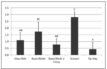
Fig. 2: Means and standard deviations of the scores of different cutting methods. Means followed by the same letter had no statistically significant difference (p>0.05).
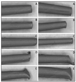
Fig. 3: Representative images of cone tips after different cutting methods: cone cut with glass slab (A); cone cut with razor/blade twice (B); cone cut with TipSnip (C); cone cut with Razor/Blade once (D); cone cut with scissors (E).
DISCUSSION
Three-dimensional sealing of root canals is important in achieving endodontic success1,3,7,10,24. The sealing ability of filling materials and the fit of the main gutta-percha cone in the apical foramen are essential to obtain this proper sealing5,25. The gutta-percha cone cut should allow a good fit with no irregularities on the final surface obtained. Discrepancies at the cone tip after cutting can theoretically prevent the fit required in the apical third, allowing infiltration8,15,22. There are several methods for cutting gutta-percha cones, usually with the aid of a device to calibrate them. Among the commercially available devices is the gauge, which needs an auxiliary tool to make the calibrated cone cut. The most commonly used cutting tools are scissors, scalpel blades and razor blades8,14,22. A new device called TipSnip both calibrates and cuts the cone. The results of this study indicate that TipSnip is the best cutting method for obtaining a regular surface on the gutta-percha cone. Some observations can be made on the samples, such as the rounding of the cone surface in the area where the cut was started while the opposite surface, where the cut is finished, is straight. However, the apical surface of gutta-percha shows no irregularities. A disadvantage of this new method is the additional cost of the device. Similar results were observed in the group in which the cone was cut in two strokes with a scalpel blade, where the irregularities formed with the first cut are removed by the second stroke, providing a satisfactory regular surface.
Studies have shown that using a razor blade against a hard surface such as a glass slab is a good method for cutting gutta-percha8,22. However, it does not allow accurate calibration of the auxiliary cone. Therefore, the method of measuring the area to be cut before cutting on the glass slab has been included in this study. Measuring the cone and using a glass slab to cut on also led to satisfactory results in the regularity of the cone surface, whereas measuring and marking the surplus for cutting hinders the accuracy of the cut because the operator might mark the cutting surface incorrectly or inaccurately. The single cut with the scalpel blade against the gauge led to the formation of an irregular cone surface that was consistently observed in the samples. It was found that the cones were rendered irregular when the scalpel blade reached the end of the cut, by a surplus of gutta-percha retained between the cutting surface of the blade and the gauge. The samples cut with scissors have the most noticeable irregularities on the surface of the cones, with the formation of two planes converging in apical direction. Similar results have been reported, contraindicating the use of this method to cut the cones.
Because of the importance of getting a good guttapercha apical fit, the results of this study lead to the conclusion that regular gutta-percha surfaces can be obtained by different cutting methods, such as the TipSnip, the cut with a double-blade and the use of a razor blade against glass slab. Thus, the professional should select the best method considering results, execution time, practicality and cost.
1. Schilder H. Filling root canals in three dimensions. Dent Clin North Am 1967;11:723-744. [ Links ]
2. Venturi M. An ex vivo evaluation of a gutta-percha filling technique when used with two endodontic sealers: analysis of the filling of main and lateral canals. J Endod 2008;34:1105-1110. [ Links ]
3. Aguiar CM, Camara AC, Araujo DSC, Santiago IMA. Estudo comparativo do selamento apical de diferentes cones de guta-percha. Rev Cienc Odontol Bras 2007;10:32-36. [ Links ]
4. Sundqvist G, Figdor D, Persson S, Sjogren U. Microbiologic analysis of teeth with failed endodontic treatment and the outcome of conservative re-treatment. Oral Surg Oral Med Oral Pathol Oral Radiol Endod 1998;85:86-93. [ Links ]
5. Santos J, Tjaderhane L, Ferraz C, Zaia A, Alves M, De Goes M, Carrilho M. Long-term sealing ability of resin-based root canal fillings. Int Endod J 2010;43:455-460. [ Links ]
6. Gurgel-Filho ED, Andrade Feitosa JP, Teixeira FB, Monteiro de Paula RC, Araujo Silva JB, Souza-Filho FJ. Chemical and X-ray analyses of five brands of dental guttapercha cone. Int Endod J 2003;36:302-307. [ Links ]
7. Pinheiro CR, Guinesi AS, de Camargo EJ, Pizzolitto AC, Filho IB. Bacterial leakage evaluation of root canals filled with different endodontic sealers. Oral Surg Oral Med Oral Pathol Oral Radiol Endod. 2009;108:e56-60. [ Links ]
8. Lopes HP, Siqueira-Jr JF, Elias CN. Scanning electron microscopic investigation of the surface of gutta-percha cones after cutting. J Endod 2000;26:418-420. [ Links ]
9. Alisson DA, Michelich RJ, Walton RE. The influence of master cone adaptation on the quality of the apical seal. J Endod 1981;7:61-65. [ Links ]
10. Brosco VH, Bernardineli N, Moraes IG. “In Vitro” evaluation of the apical sealing of root canals obturated with different techniques. J Appl Oral Sci 2003;11:181-185.
11. Haikel Y, Wittenmeyer W, Bateman G, Bentaleb A, Allemann C. A new method for the quantitative analysis of endodontic microleakage. J Endod 1999;25:172-177. [ Links ]
12. De Deus QD. Endodontia. 5a edicao. Rio de Janeiro: Medsi, 1992. [ Links ]
13. Moule AJ, Kellaway R, Clarkson R, Rowell J, MacFarlane R, Lewis D, et al.Variability of master gutta-percha cones. Aust Endod J 2002;28:38-43. [ Links ]
14. Araujo Filho WR. Analise in vitro do selamento marginal apical de duas tecnicas de obturacoes de canais radiculares associadas a dois tipos de cones mestres. Rev Bras Odontol 2007;64:76-79. [ Links ]
15. De-Deus G, Maniglia-Ferreira CM, Gurgel-Filho ED, Paciornik S, Machado AC, Coutinho-Filho T. Comparison of the percentage of gutta-percha-filled area obtained by Thermafil and System B. Aust Endod J 2007;33:55-61. [ Links ]
16. Kerekes K. Evaluation of standardized root canal instruments and obturating points. J Endodon 1979;5:145-150. [ Links ]
17. Mayne JR, Shapiro S, Abramson II. An evaluation of guttapercha points.1. Reliability and validity of standardization. Oral Surg Oral Med Oral Pathol 1971;31:250-257. [ Links ]
18. Goldberg F, Gurfinkel J, Spielberg C. Microscopic study of standardized gutta-percha points. Oral Surg Oral Med Oral Pathol 1979;47:275-276. [ Links ]
19. Allison DA, Michelich RJ, Walton RE. The influence of master cone adaptation on the quality of the apical seal. J Endod 1981;7:61-65. [ Links ]
20. Inan U, Aydin C, Tunca YM, Basak F. In vitro evaluation of matched-taper single-cone obturation with a fluid filtration method. J Can Dent Assoc 2009;75:123. [ Links ]
21. Lopes HP, Siqueira JF Jr. Endodontia. Rio de Janeiro, Brazil, Ed Medsi, 2010. [ Links ]
22. Jacobsen EL. Clinical aid: adapting the master gutta-percha cone for apical snugness. J Endod 1984;10:274. [ Links ]
23. Gallin DM. Changes observed upon cutting the gutta-percha points. NY State Dent J 1984;50:212-213. [ Links ]
24. Wolcott J, Himel VT, Powell W, Penney J. Effect of two obturation techniques on the filling of lateral canals and the main canal. J Endodon 1997;23:632-635. [ Links ]
25. Nagas E, Altundasar E, Serper A. The effect of master point taper on bond strength and apical sealing ability of different root canal sealers. Oral Surg Oral Med Oral Pathol Oral Radiol Endod 2009;107:e61-64. [ Links ]

