


Services on Demand
Journal

Article
 Spanish (pdf)
 Article in xml format
 Article references
 Send this article by e-mail
Indicators
-
 Cited by SciELO
Related links
-
 Similars in
SciELO
Similars in
SciELO
Share

 Permalink
Revista veterinaria
On-line version ISSN 1669-6840
Rev. vet. vol.24 no.2 Corrientes Dec. 2013
TRABAJOS DE INVESTIGACIÓN
Accidental poisoning with Wedelia glauca ("sunchillo") in a bull confirmed by analysis of rumen content
Costa, E.1; Zeinsteger, P.1; Streitenberger, N.1; Gimeno, E.2; Fazzio, L.1
1 Facultad de Ciencias Veterinarias, Universidad Nacional de La Plata, calle 60 y 118, CC 296, B1900AVW, La Plata, Argentina. E-mail: pzeins@fcv.unlp.edu.ar
2 Consejo Nacional de Investigaciones Cient. y Técnicas (CONICET), Argentina.
Abstract
Wedelia glauca is an invasive, perennial plant of the Asteraceae family native to South America. Its toxicity is attributed to the presence of a hepatotoxic terpenoid known as atractyloside, a powerful inhibitor of cellular respiration and ATP synthesis. Cattle are the most frequently poisoned species, and the course of this poisoning is hyperacute or acute. Occasionally, it is possible to fnd fragments of plants in the rumen contents and indentify the dermis structure of the plants, as they do not undergo significant changes in spite of the mechanic and enzymatic activities occurring in the rumen. The macroscopic and microscopic anatomopathologic findings of a natural Wedelia glauca poisoning case in a Hereford bull are reported. It was confirmed by micrographic analysis of plant fragments found in the rumen contents and also in bales used to feed those animals.
Key words: Bull; Poisoning; Wedelia glauca; Micrographic analysis.
Intoxicación accidental de un toro con Wedelia glauca ("sunchillo") confirmada por análisis de contenido ruminal.
Resumen
Wedelia glauca es una planta perenne e invasiva perteneciente a la familia Asteraceae que se encuentra en América del Sur. Su toxicidad se debe a la presencia de un terpenoide hepatotóxico conocido como atractilósido, un potente inhibidor de la respiración celular y de la síntesis de ATP. El ganado bovino resulta intoxicado con frecuencia, siendo el curso de la afección hiperagudo o agudo. Ocasionalmente es posible encontrar fragmentos vegetales en el contenido ruminal y así identificar la estructura de la dermis de las plantas, ya que no sufren cambios significativos a pesar de los procesos mecánicos y enzimáticos del rumen. Se presentan los hallazgos macroscópicos y microscópicos de un caso de intoxicación natural por Wedelia glauca en un toro Hereford, confirmado por el análisis micrográfico de fragmentos vegetales hallados en contenido ruminal y fardos de alfalfa utilizados en su alimentación.
Palabras clave: Toro; Intoxicación; Wedelia glauca; Análisis micrográfico.
Recibido: 14 agosto 2013
Aceptado: 20 septiembre 2013
INTRODUCTION
Wedelia glauca (Wg) [Wedelia glauca (Ort.) Hoffm. ex. Hicken] belongs to the Asteraceae family which comprises a great deal of specimens, some of them being well-known as poisoning species to human beings and domestic animals 16, 20. Poisonous plants have associated toxins which constitute the plants' chemical defense mechanism against insects or herbivores 13, 15.
Wg is a widespread plant in South America, especially in Argentina, Chile, Paraguay, Southern Brazil and Uruguay 4, 6, 11, 17, 20. It is a perennial and herbaceous plant, 30 to 80 centimeters high, with stiff stems and simple or compound lanceolate basal leaves with 2-3 teeth (Figure 1). The yellow inflorescences are arranged in terminal capitates heads 4, 5, 18 (Figure 2). Fructification occurs during summer and at the beginning of autumn. Only the underground parts of the plants remain alive the rest of the year. Wg toxicity is attributed to the presence of a hepatotoxic terpenoid called atractyloside, a powerful inhibitor of cellular respiration and ATP synthesis 7.
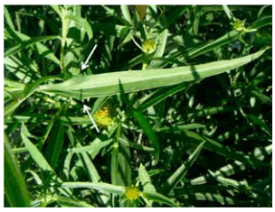
Figure 1. Leaves of Wedelia glauca. Basal teeth are shown with arrows.
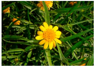
Figure 2. Inflorescence of Wedelia glauca.
The atractyloside inhibits the ADP/ATP carrier through the membrane of the organelles, preventing the oxidative phosphorylation by blocking the translocation of adenine dinucleotide 12. The consequence is an initial alteration of the intrahepatic circulation with hepatocyte necrosis and periacinal haemorrhage (centrilobular). Such findings are common to acute hepatotoxic compounds 6, 19.
Deaths attributed to Wg poisoning in domestic animals occurs frequently 17. Most of the cases are due to the presence of Wg in the forage, mainly in alfalfa bales contaminated with the weed. Cattle are the most frequently poisoned species. Cases in other species such as llama, giraffe, axis, deer and mufouns have occasionally been reported in zoos 6, 8. The course in cattle, sheep and horses ranges from hyperacute to acute, developing between 2 and 46 hours. Although Wg poisoning shows characteristic anatomopathological findings, sometimes complementary methods to confirm the diagnosis are necessary, mainly when the number of dead animals is large or when the economic value of a particular animal is high.
Sometimes, it is possible to find fragments of plants in the rumen content 21. The dermis of the plants does not undergo significant changes in spite of the mechanic and enzymatic activity occurring in the rumen. This allows the identification of such structures by means of optical microscopy, a method known as micrography or micrographic analysis.
The macroscopic and microscopic anatomopathologic findings of an accidental poisoning case with Wg confirmed by micrographic analysis of plant parts found in the rumen contents and the bales used to feed animals are presented in this paper.
MATERIAL AND METHODS
Case history. In an artificial insemination centre in Buenos Aires, Argentina, a-three-years-and-a-half Hereford bull was found dead, and no signs or previous disease was report by the owner. The farm had 60 bulls distributed in individual pens for semen extraction. Feeding was based on oat concentrate, alfalfa bales and vitamin-mineral nucleus, administered twice a day (08:30 AM and 04:30 PM.). The rest of the bulls showed no clinical signs, changes in behavior or feeding intake.
Necropsy and tissue samples. General necropsy of the bull was performed, and samples from liver, kidney, heart and lungs were gathered for microscopic evaluation. The histological samples were fixed in 10% neutral buffered formalin, embedded in paraffin, sectioned, and stained with hematoxylin and eosin.
Micrographic analysis. As death was suspected to be caused by a poisonous plant, samples of rumen contents from different strata, as well as a contaminating whole plant collected from one alfalfa bale, were taken for further analysis and kept in labeled polyethylene bags. Both rumen contents and leaves of the poisonous plant were washed with tap water and then submitted to diaphanisation in a glass beaker, adding 10 ml 5% NaOH solution and heating for about 10 minutes, in order to eliminate the excess of chlorophyll that may interfere with further microscopic observation. The alkaline solution was discharged by washing plant materials for 4-6 times with distilled water. Later, fragments from rumen contents and leaves from the plant were mounted separately on slides with cover slips. All the observations were registered with a digital camera mounted on a microscope.
RESULTS
Examination of the pen showed signs of pedaling where the animal succumbed. External examination of the bull showed abundant foam from nostrils and red liquid leaking from nostrils and anus.
Gross lesion. The necropsy revealed abomasal congestion, petechial hemorrhages and mucosal suffusions. In the duodenum and the first portion of the jejunum the content was red and petechial hemorrhages in the mucosa were verified. Liver was swollen and with dark red mottling on the cut surface. In the gallbladder petechial hemorrhages were observed on the serous membrane. When the rumen was opened, a large amount of food and leaves which had not been digested were found (Figure 3). Other findings included petechial hemorrhages in epicardium and endocardium and congestion of the lungs; all other organs appeared normal. The pH in the rumen content was 6 and tests for the determination of cyanhydric acid and nitrites or nitrates were negative.
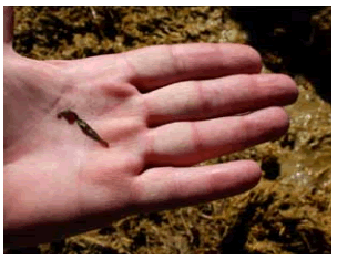
Figure 3. Plant material found in the ruminal content of the bull.
Microscopic study. Histopathology revealed intense congestion of the liver and, in periacinar zone the hemorrhage was more evident. The nuclei of hepatocytes in such areas were pyknotic while other cells showed karyolisis or karyorrhexis, as well as evident cytoplasmic fragmentation, all indicating severe coagulative necrosis.
Micrographic analysis. Micrographic observation of the plants obtained from the rumen (leaves) and one of the alfalfa bales -whole plant- showed long polyhedral epidermal cells with rounded edges surrounded by 3-4 anomocytic and anisocytic stomata (Figure 4) A type of tector trichome prevailed, with a long body formed by several cells and a triangular apical cell; the base of these trichomes showed a rosette-like structure formed by specialized epidermal cells (Figure 5). Some of the trichomes showed ornamentation.
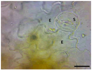
Figure 4. Epidermal cells (E) and stomata (S) of Wg. Treatment with NaOH 5% at 100°C. Scale: 50 mm.
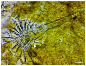
Figure 5. Tector trichome (T) and epidermal cells in rosette-like structure (R). Treatment with NaOH 5% at 100°C. Scale: 50 mm.
DISCUSSION
Poisonous plants usually have defense mechanisms against herbivores; either they have a disgusting taste and smell, or they are astringent, coriasea or bristly 2, 3. Despite these botanical features, animals eat them under extreme hunger or when the plants are mixed in the forage, affecting their health in many ways.
As in many countries with weather conditions ranging from warm to sub-tropical climates, Argentina is environmentally suitable for extensive cattle production, mainly in the central and north-east regions of the country. Almost 45 million heads are bred only in these two areas 14. Poisonous weeds are also widespread in such regions, and considering its geographic extension their presence is a permanent concern for livestock farmers.
The intake of poisoning plants can also affect a large number of grazing animals. Even in intensive breeding systems, like the feed-lots, poisonous plants can produce high mortality. Low quality hay used to provide feed-lots with fiber constitutes a health risk and can be a potential problem if it is mixed with poisonous plants 9. Occasionally, only one animal gets poisoned, usually as a result of consuming food accidentally contaminated with a poisonous weed.
For the case presented here, clinical signs as well as macro and microscopic analysis direct the diagnosis to acute liver poisoning. However, a wide variety of agents produce similar damages. Wg, Cestrum parqui, Xanthium spp., Mioporum laetum and some hepatotoxic compounds such as copper are the most commonly diagnosed in Argentina. Sometimes it is important to acknowledge the etiological agent in cases of death, and the micrographic study of the rumen content is usually an important tool to achieve this aim.
Micrographic analysis is a simple method used for different purposes, e.g. botanic identification of plants. Regarding animal biology, it is considered a useful tool to determine the nutritional behavior of wild and domestic animals 1, 10. It is important to highlight that digestion does not alter the features of trichomes, stomata and even epidermal cells of forages.
As significant alterations were not produced in such structures, it was possible to confirm the ingestion of a poisonous plant by means of micrographic analysis. The technique is simple and inexpensive, but strong knowledge regarding the anatomy of plants is necessary. Besides, a data base that includes the records of poisonous plants present in a specific geographical area is necessary. If these conditions are met, micrographic analysis can be used as a complementary method to confirm Wg poisoning, as well as other poisonous plants.
1. Alipayo D, Valdez R, Holechek JL, Cardenas M. 1992. Evaluation of microhistological analysis for determining ruminant diet botanical composition. J Range Manage 45: 148-152. [ Links ]
2. Bruce TJ, Pickett JA. 2007. Plant defence signalling induced by biotic attacks. Current Opin Plant Biol 10: 387-392. [ Links ]
3. Bruneton J. 2001. Plantas tóxicas. Vegetales tóxicos para el hombre y los animales, 2ª ed., Acribia, Zaragoza, 540 p. [ Links ]
4. Fernández Pazos C. 2010. Alerta, sunchillo (Wedelia glauca). Rev Brangus 32: 66-72. [ Links ]
5. Giannitti F, Margineda CA, Cid MS, Diab SS, Weber N, Rodríguez A, Campero CM, Odriozola ER. 2012. Mortality of a captive axis deer (Axis axis) and a llama (Lama glama) due to ingestion of Wedelia glauca. J Vet Diagn Invest 24: 1068-1072. [ Links ]
6. Giannitti F, Margineda CA, Cid MS, Montobbio C, Soteras CI, Caffarena RD, Diab SS. 2013. Fatal Wedelia glauca intoxication in calves following natural exposure. Vet Pathol 50: 530-503. [ Links ]
7. Lemaster J, Sowers A. 1979. Phosphate dependence and atractyloside inhibition of mitochondial oxidative phosphorylation. J Biol Chem 254: 1248-1251. [ Links ]
8. Magnano G, Ciallella M, Sánchez E, Flores A, Riveros M, Macias A, Schleef N. 2011. Un caso de intoxicación en llamas (Lama glama) por Wedelia glauca. Vet Arg 28: 279. [ Links ]
9. Micheloud JF, Rodríguez AM, Cámpora L, Webber N, Campero CM, Odriozola ER. 2012. Caso inusual de calcinosis enzoótica por el consumo de Solanum glaucophyllum en un encierre a corral. Rev Med Vet 93: 59-62. [ Links ]
10. Morrison J, Smith S, Aiken G, Huo C. 2008. Using microhistological techniques to predict botanical composition of horse diets on cool-season grass pasture. Proceedings Joint IGC/IRC International Meetings Grassland Society (Mongolia, China). On line: http://www2.ca.uky.edu/pss/publications.php. [ Links ]
11. Mujawar I. 2013. Wedelia glauca (Ortega) O. Hofmm Ex Hicken (Asteraceae): Poisonous weed from Urun-Islampur of Sangli district of Maharashtra. Ind J Fund Appl Life Sci 3: 92-94. [ Links ]
12. Obatomi DK, Bach PH. 1998. Biochemistry and toxicology of the diterpenoid glycoside atractyloside. Food Chem Toxicol 36: 335-346. [ Links ]
13. Odriozola E. 2004. Limitantes de la producción: causas tóxicas. 2ª Jornada de Actualización Ganadera. Balcarce, Buenos Aires. On line: http://www.produccion-animal.com. ar/sanidad_intoxicaciones_metabolicos/intoxicaciones/48-limitantes_produccion_causas_toxicas.pdf. [ Links ]
14. Programa de las Naciones Unidas para el Desarrollo (PNUD). 2009. Caracterización de la producción ganadera en Argentina frente al cambio climático. On line: http://www.undp.org.ar/docs/prensa/brief-05-cambios.pdf. [ Links ]
15. Rasmann S, Agrawal AA. 2009. Plant defense against herbivory: progress in identifying synergism, redundancy, and antagonism between resistance traits. Current Opin Plant Biol 12: 473-478. [ Links ]
16. Riet-Correa F, Méndez MC, Schild AL. 1993. Intoxicações por plantas e micotoxicoses em animais domésticos, Ed. Hemisferio Sur, Montevideo, 340 p. [ Links ]
17. Rivero R, Delgado ML, Matto C, Novoa F, Uriarte G, Charbonier D. 2010. Intoxicación por Wedelia glauca en bovinos en Uruguay. Veterinaria (Montevideo) 46: 39-45. [ Links ]
18. Rodriguez Armesto R, Peralta C, Zinmerman R, Ochoteco M, Repetto A, Picco EJ. 2003. Mortandad en bovinos atribuible a la ingestión de Wedelia glauca. Vet Arg 20: 745-751. [ Links ]
19. Santos JC, Riet-Correa F, Simõs SV, Barros CS. 2008. Patogênese, sinais clínicos e patología das doenças causadas por plantas hepatotóxicas em ruminantes e eqüinos no Brasil. Pesq Vet Bras 28: 1-14. [ Links ]
20. Tokarnia CH, Brito MF, Barbosa JD, Peixoto PV, Döbereiner J. 2012. Plantas tóxicas do Brasil para animais de produção, Ed. Helianthus, Rio de Janeiro, 586 p. [ Links ]
21. Yagueddú C, Cid S, López T. 1998. Microhistological analysis of sheep gastro-intestinal content to confrm poisonous plants ingestion. J Range Manage 51: 655-660. [ Links ]
