


Services on Demand
Journal

Article
 English (pdf)
 Article in xml format
 Article references
 Send this article by e-mail
Indicators
-
 Cited by SciELO
Related links
-
 Similars in
SciELO
Similars in
SciELO
Share

 Permalink
Revista argentina de cardiología
On-line version ISSN 1850-3748
Rev. argent. cardiol. vol.83 no.3 Ciudad Autónoma de Buenos Aires June 2015
IMAGES IN CARDIOLOGY
Ventricular Perforation by Pacemaker Lead
Perforación ventricular por catéter de marcapasos
MIGUEL RUBIOMTSAC, MARÍA L. SÉSTITO, JULIO BALDI (H)
The images correspond to a 70-year-old female pa-tient with permanent DDD pacemaker implanta-tion in July 2012, without complications during the procedure and with good immediate device performance. Two years later, the patient underwent mi-tral valve replacement as a result of severe chronic mitral valve disease. Median sternotomy surgery revealed multiple loose adhesions and ground-glass opacity in the epicardium, and the presence of the distal end of the ventricular pacing lead in the pericardial cavity, perforating the right ventricular inferior wall (Figure 1). Review of the chest X-ray following pacemaker implantation indicated that the tip of the ventricular lead was placed in the pericardial cavity (Figure 2).
Conficts of interest
None Declared
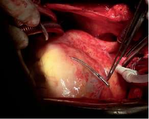
Fig. 1.

Fig. 2.
Clínica Bazterrica, Buenos Aires, Argentina
MTSAC Full Member of the Argentine Society of Cardiology

