


 English (pdf)
 Article in xml format
 Article references
 Send this article by e-mail
 Cited by SciELO
  Similars in
SciELO
Similars in
SciELO 
 Permalink
CASE REPORT
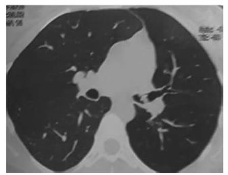
22-year-old female patient with history of bronchial asthma since childhood. The patient was referred to the bronchoscopy service to be evaluated for suspected endoluminal tumor. She brings a normal spirometry. The chest tomography shows endoluminal lesion at the left main bronchus (LMB). The tomography also shows volume reduction of the left lung field with homolateral mediastinal laterality and herniation of the right lung towards the left. The physical examination shows generalized sibilance in the left lung field, without any other alteration.
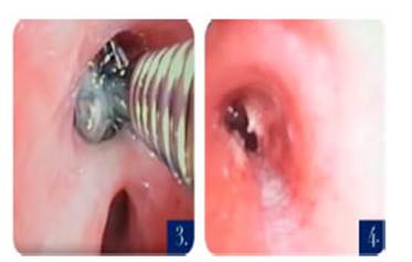
We performed a fibrobronchoscopy where we observed a thick division spur 4 cm away from the base of the left main bronchus (LMB). At first we thought it was the bronchus spur that divides the upper and lower lobe, but immediately after such “spur” we saw a rounded, off-white, wrinkled, mobile lesion. It is assumed that the image interpreted as a spur could be an adhesion located at the distal level of the LMB that doesn’t allow access to bronchial segmentation or the removal of the mobile lesion that we observed. We performed radiofrequency ablation at the middle part of the adhesion. After cutting the adhesion, it was possible to access the normal bronchial segmentation and remove with a biopsy bracket the off-white formation that was covered with fibrin and desiccated mucus secretions. When we analyzed it outside the patient, after removing the layer of secretions, we could identify the cap of a pen.

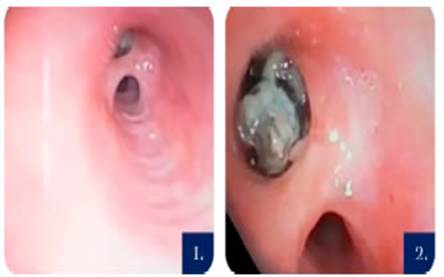
1. “Division spur”, 4 cm away from the LMB. 2. Off-white lesion through the spur. 2. Off-white lesion through the spur.
The patient remembered that when she was 5 years old, she was playing with a pen and aspirated the cap. Our patient has a favorable evolution, she no longer needs bronchodilator treatment because she is better and has no symptoms.
DISCUSSION
The aspiration of a foreign body is an uncommon clinical entity in adults and requires a high index of clinical suspicion for its diagnosis, especially in people without a history of foreign body aspiration or without the presence of any of the risk factors, such as advanced age, use of sedatives, neurological or neuromuscular disorders, traumatisms, alcoholism or handling of tracheostomy cannulas1-3.
Foreign bodies can be classified in organic and inorganic substances; the aspiration of the latter is common in children and young people who introduce the substance into their mouth as entertainment1.
A chest tomography is recommended to see the location and size of the foreign body in the airway, being the tomography more sensitive and specific for the diagnosis2-4.
Late diagnosis of the aspiration of a foreign body in the airways causes chronic respiratory symp toms, namely sibilance, dyspnea, and recurring infections, which are commonly confused with other respiratory diseases such as asthma, among others. Thus, wrong treatments are indicated without any improvement1-3,6.
When a foreign body remains in the airway for long periods of time, it produces an inflammatory reaction localized in the airway that can result in the appearance of granulation tissue due to inflam mation and hyperplasia of the adjacent bronchial mucosa and is present the whole time between the aspiration event and the removal of the foreign body, originating anatomical alterations that are visible through bronchoscopy4,5.
Flexible bronchoscopy is safe, it allows for a more complete study of the airways and has a high rate of success in the identification and removal of foreign bodies1-4.

