


 English (pdf)
 Article in xml format
 Article references
 Send this article by e-mail
 Cited by SciELO
  Similars in
SciELO
Similars in
SciELO 
 Permalink
I Introduction
Bacterial biolms are clusters of bacteria that are attached to a surface and/or to each other and embedded in a self-produced matrix 1. The biolm matrix consists of extracellular polysaccharides (EPS), proteins, glycopeptides, nucleic acids, and lipids 2. Compared with planktonic forms, organisms in biolms resist undesirable physical, chemical and biological factors in the environment, host immune system, and antimicrobial therapy 3. Therefore, the susceptibility of bacteria in biolms to antimicrobial agents is 500{5000 times lower than that of bacteria in suspension (planktonic) cells of the same microorganism 4. This resistance is due to restricted penetration of the biolm matrix, the presence of antimicrobial enzymes, an altered growth rate inside the biolm, a stress response to unfavorable environmental conditions, and over-expression of genes 5.
The detrimental impacts of bacterial adhesions generate serious environmental problems, public health risks and, eventually, vast economic losses. Environmentally, biolm in association with algae and other organisms disrupts the natural ecosystem by causing biofouling, microfouling, macrofouling and inorganic fouling, which ultimately aects water quality, causing alterations in taste, color, and odor 6. Industrially, biolm is responsible for biocorrosion of metal pipelines, blockage of ltration systems, oil spoilage, low durability of construction materials 7, and spoilage of food and dairy products 8. Medically, biolms can colonize medical devices such as prosthetic joint replacements and heart valves, pacemakers, intra-ventricular cardiac assist devices, urinary tract catheters, peritoneal dialysis catheters, central venous catheters, neurovascular shunts, synthetic vascular grafts and stents, articial voice prostheses, and intrauterine devices 9. It has been estimated that two-thirds of human bacterial infections may involve biolms 6.
A combined treatment based on applying direct electric current (DC) along with low doses of antibiotics can increase the effcacy of antibiotics on biolms: this is termed the bioelectric eect 10. DC voltage generates radicals as a result of electrolysis of the medium, which is suggested as a principal factor in its eectiveness 11. In addition, some reports describe enhanced effcacy due to improved antibiotic binding to biolms 12, and enhanced biolm detachment , since an external DC electrostatic force can increase the area of bacterial exposure to the antibiotics 13. The use of DC without antibacterial agents to reduce biolm formation may prevent biolm formation on certain biotic and abiotic substrates. DC can increase the repulsive electrostatic forces between organisms and the adhesive surface 14.
In addition, DC can reduce biolm formation by changing physical conditions (e.g., temperature, pH) at the adhesive surface, and through the accumulation of products of oxidative stress 15. Previous studies have demonstrated that DC exhibits bactericidal activity against established biolms 16, 17. This has the potential benet of eliminating the use of traditional antimicrobials, thus decreasing the risk of selective resistance to these agents 18.
Nanoparticles (NPs) exhibit excellent antimicrobial activities 19, 20 and are already being used in many commercial products, including toothpaste, sunscreen, and food products 21, 22. NPs are considered a promising tool for the treatment of bacterial biolms because antibiotic resistance mechanisms are not eective against them. NPs can enter a biolm system, settle on its surfaces and migrate to its inner portion 23. They then interact with microbes and EPS and can subsequently reduce microbial activities and alter population structure pollutants 24. Of all the NPs, the most promising and widely studied are metal oxides, such as TiO2 25. TiO2-NPs have excellent antimicrobial activity and constitute one of the most extensively manufactured and used nanomaterials, with a global production of 5500 tons per year 26. TiO2-NPs are commonly used in paints, pigments, food, cosmetics, coatings, paper, catalysts, and plastics 27, 28. They can inhibit bacterial growth and biolm formation 29, and have excellent antimicrobial eects on biolm formation and the chronic toxicity of mature biolms 30. However, the continuous release of metal oxide ions such as TiO2 from TiO2-NPs causes massive production of oxygen free radicals or reactive oxygen species (ROS). The smaller particle size of TiO2-NPs allows them to pass through the EPS matrix of the biolm and enter the cells through porins, waterlled channels that aid the process of exchange/transport of low molecular weight compounds with the ambient environment. When NPs enter the cytoplasm they have an even more destructive eect on metabolism and biochemical activities, particularly respiration and subsequent energy-dependent cellular processes 31. NPs bind mainly to -SH groups of amino acids and formations of extra -S-S- bonds. The resulting conformational changes in protein structure lead to protein inactivation and ribosome denaturation 32.
In this way enzymes in the respiratory chain are deactivated; this is followed by the obstruction of electron transport by oxygen and nally by blockage of ADP-phosphorylation to ATP. At a genetic level, TiO2-NPs bind to nucleic acids, blocking DNA replication and repair processes 33. Also reported has been a lower ability of P. aeruginosa PAO1 to assimilate and transport iron and phosphorous, and inhibition of the biosynthesis and degradation of heme (Fe-S cluster) groups 34. The present study aims to compare the eect ofdierent TiO2-NP concentrations with the eect of dierent low DC conditions on the biolms of Bacillus cereus and Pseudomonas aeruginosa as models of Gram-positive and Gram-negative bacterial biolms.
II Materials and methods
i TiO2-NPs
The TiO2-NPs (Aeroxide® TiO2 P25) were purchased from Sigma Aldrich Inc., USA. TiO2 is composed of anatase (85%) and rutile (15%) crystal structure; the mean diameter of the Nano-TiO2 particles is 21 nm. Nanoparticles were supplied as white nano-powder of hydrophilic fumed titanium dioxide nano-particles.
ii Characterization of TiO2-NPs
TiO2-NPs were characterized by TEM, SEM, XRD, and FTIR 35, 36. The surface morphology and diameter of the nanoparticles were measured using TEM and SEM (ZEISS, Germany). The Xray diraction (XRD) patterns of the samples were characterized using an X-ray diractometer with Cu-K radiation. The crystalline nature of the TiO2-NPs was recorded using X-ray diraction (XRD) (Bruker, Germany) with CuK radiation (1.5406 A) in the 2scan range of 10-90°. The FTIR spectrum of TiO2-NPs was recorded on Fourier Transform Infrared Spectrophotometer (Bruker, Germany) in the region of 4000 to 500 cm-.
iii DC exposure system
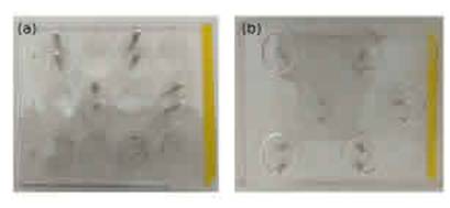
A DC power supply (Etommens eTM-305A, made in China) was used to deliver 9 V, 6 mA direct electric current. The electric current was applied through a pair of silver/silver chloride electrodes. The anode is designed in the form of two connected pieces. The lower part is a circle of 1 cm radius, while the upper part is a long plate. The diameter of the circle part is designed to t the diameter of the well of the 12-well polystyrene microtitre. The cathode is a long rod plate placed at a distance of 1 cm from the anode. Both cathode and anode rods were xed to two holes in the lid of a cover of the microtitre plate. These electrodes were repeated to face each well in the microtitre plate. Figure 1 illustrates an image of the electrodes: Fig. 1(a) is an overhead view while Fig. 1(b) is an inverted view.
iv Antibiolm potential of TiO2-NPs and DC in the eradication of established biolm
a Biolm Formation
B. cereus and P. aeruginosa were examined as models for Gram-positive and Gram-negative bacteria, respectively. Initially, 3 mL of tryptone soy broth (TSB), supplemented with 1% glucose-containing 108 CFU/mL (0.5 McFarland) of each bacterial culture, was put into a 12-well polystyrene microtitre plate, and TSB was incorporated as a negative control. The plate was incubated for 24 h under aerobic static incubation at 37 - to allow the formation of a multilayer biolm. After incubation, the planktonic non-adherent cells were blotted out and each well washed three successive times with 3 ml of physiological saline solution.
b Application of TiO2-NPs and DC to the biolms
The preformed biolms were suspended in 3 mL of fresh TSB and exposed to dierent treatments (electricity or TiO2-NPs). In the TiO2-NP exposure groups 200 uL of various concentrations of TiO2-NPs, ranging from 5 to 160 ug/mL, were added to each well and incubated as mentioned previously. In the DC exposure groups DC was applied to three groups of each biolm type for 5, 10, and 15 min respectively. The electrical energies used were therefore 16.2 J, 32.4 J, and 48.6 J, respectively. Temperature was measured every 5 min using a digital thermometer. After incubation the adhered bacterial slimes were quantied using 0.3% crystal violet (CV) solution. The dye attached to the surface-adhered cells was solubilized with acetic acid (33%) and determined spectrophotometrically at 590 nm (Tecan Innite M200, Switzerland). The eradication percentage of the biolms was calculated by the following equation 37:
Where A represents the absorbance of the untreated control wells and A0 the absorbance of the treated wells. All the experiments were performed in triplicate, and the following methods were used to evaluate the eect of each protocol applied to each biolm type.
v Exopolysaccharide (EPS) and protein content assessment
For determination of the exopolysaccharide (EPS) content of detached biolms, the decanted cell-free supernatant was added to three volumes of ice-cold absolute ethanol and incubated overnight at 4 C°. The resulting pellets were centrifuged, dried and their yield was assessed using the phenol-sulfuric acid approach. Brie y, 0.1 mL of EPS samples were mixed with 1.0 mL of cold phenol (6 %) and 5.0 mL of sulfuric acid 95 % (v/v); the mixture was shaken and incubated for 10 min. Absorbance was measured at 490 nm. The EPS content of each sample was calculated using the glucose standard curve 38. Protein content was also determined 39. Based on the Bradford method, we mixed well 100 L of detached biolms with 5 mL Bradford solution (100 mg Coomassie Brilliant Blue G- 250 was dissolved in 50 mL 95% ethanol, then 100 mL of 85% phosphoric acid (H3PO4) was carefully mixed in by stirring, completing to 1 L total volume). After 5 min incubation at room temperature, absorbance was measured at 595 nm. A standard curve was constructed by BSA (0, 0.0625, 0.125, 0.25, 0.5 and 1 g/L).
vi Cell surface hydrophobicity evaluation
The cell surface hydrophobicity (CSH) of the bacterial cultures, control and treated, was determined. The bacterial cells in detached biolms were harvested by centrifugation at 12000 rpm for 10 min, and the pellets obtained washed twice with sterile PBS. The bacterial suspensions were vortexed vigorously with an equal volume of hydrocarbon (e.g., xylene), held at room temperature for 5 min. The absorbance of the aqueous phase was determined spectrophotometrically at 600 nm (Labomed. model UV-Vis Double beam spectrophotometer) 40.
vii Reactive oxygen species assay
The reactive oxygen species (ROS) produced by the bacterial cells as a result of the dierent treatments with electricity and NPs were evaluated by FDA, 3,6-diacetoxy uoran assay. Brie y, 100 uL of FDA (10 ug/mL) was added to each treated sample and control, then incubated for 30 min at 30 C°. After incubation, cleavage of the FDA was stopped by the addition of acetone to a nal concentration of 50% v/v. To eliminate suspended particles, the mixture was centrifuged for 5 min at 10000 rpm. Fluorescence intensity was analyzed by a uorimeter microplate reader (FluoStar Omega, Germany) with excitation and an emission wavelength of 495 nm and 525 nm, respectively. ROS concentration was determined by the standard curve of H2O2 at dierent concentrations 41.
viii Study of morphological changes in biolm using SEM
The in uence of dierent treatments on biolm disintegration was visualized by scanning electron microscopy. Biolms were grown on glass coverslips (11 mm) submerged in a 12-well polystyrene microtitre plate containing 108 CFU/mL both in the control and treated wells. After incubation, the coverslips were gently washed with 0.85% NaCl to remove planktonic cells. Samples were xed in 2.5% buered glutaraldehyde for 24 h, followed by washing with 4% OsO4 in 0.1 M phosphate buer for 2 h 42. Samples were dehydrated with a gradient acetone series (35-100%) for 15 min. The dried biolms were coated with gold and visualized under SEM (JEOL JSM 6360LA, Japan).
ix Statistical analysis
All data were expressed as the mean standard deviation (SD) of three independent repeats. ANOVA was used to evaluate the dierence between multiple groups. Signicant dierences between experimental groups were determined using a two-tailed Student's t-test (Excel 2013 Microsoft, USA). Results were considered statistically signicant when the p-value < 0:05.
III Results
i Characterization of TiO2-NPs
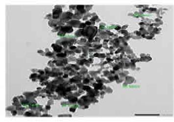
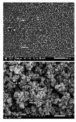
The transmission electron micrographs of TiO2- NPs in Fig. 2 show the relatively narrow dispersion characteristic and spherical morphology of NPs with diameters of 22-34 nm. The morphology and size of the NPs were characterized using scanning electron microscopy (SEM). Agglomerating and roughly spherical NPs are illustrated using SEM images in Fig. 3.
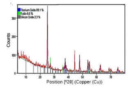
To conrm the presence of TiO2 and analyze the structure, we used a powdered sample and a CuK - X-Ray Diractometer: results are shown in Fig. 4. The peaks appeared at 2 value ranging the diraction peak at 2 with 25°, 38°, 48°, 54°, 62°, corresponds to the crystal planes of (101), (004), (200), (105) and (204) respectively, indicating the formation of the anatase phase of TiO2 43. FTIR analysis was used to determine the functional groups of TiO2-NPs. Figure 5 shows the FTIR spectrum of TiO2-NPs, in which the peaks at 3351.69 cm sired phase, so the peaks at 433.32 cm.
ii Antibiolm potential of TiO2-NPs and DC for eradication of established biofilm
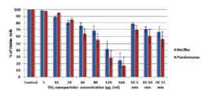
Figure 5: The eect of TiO2-NP concentrations (5-160 ug/mL) and DC exposure (5, 10, and 15 min) on the percentage of viable cells of B. cereus and P. aeruginosa biolms.
Firstly, the eect of both treatments in the eradication of preformed biolms was determined as shown in Fig. 6. The results indicate a signicant reduction in biolm adherence due to either TiO2-NPs or DC. The eradication potency increases signicantly as TiO2-NP concentrations increase, showing dose-dependent behavior. The antibiolm activity caused by TiO2-NPs ranged from 1% at 5 ug/mL to 75% at 160 ug/mL in the case of B. cereus, whereas at the same concentrations in P. aeruginosa biolm it ranged from 2% to 83% . The LD50 calculated for B. cereus is 104-4 uyg/mL while for P. aeruginosa it is 63-3 ug/mL. Regarding DC energy, as the DC exposure time (energy) increased, the disintegration percent signicantly increased. It reached 21%, 29%, and 33% at exposure time 5-, 10-, and 15-min, respectively for B. cereus and 30%, 39%, and 44% respectively for P. aeruginosa. The eect of DC exposure for 15 min is thus considered equivalent to the eect of 40 ug/mL TiO2-NPs. In addition, the EPS and protein concentrations in the decanted bacterial cultures increased under both treatments, re ecting biolm destabilization and detachment. The results of EPS and protein concentrations support the results of the eradication percentage, as shown in Figs. 7 and 8. Notably, compared to the control treatment there was a small increase of 1.2-0.2 - in temperature after 15 min of DC exposure. Temperature change is therefore not an eective factor in this work.
Figure 6: The eect of TiO2-NP concentrations (5-160 ug/mL) and DC exposure (5, 10, and 15 min) on the percentage of viable cells of B. cereus and P. aeruginosa biolms.

Figure 7: The eect of TiO2-NP concentrations (5-160 ug/mL) and DC exposure (5, 10, and 15 min) on the EPS concentration (ug/mL) of B. cereus and P. aeruginosa biolms.
iii Cell surface hydrophobicity (CSH) evaluation
Bacterial treatments that exhibited a CSH % lower than 30% were deemed hydrophilic, and those with CSH higher than 70% were considered hydrophobic; samples with CSH between 30% and 70% were categorized as moderately hydrophobic 45. As shown in Fig. 9, control biolms and biolms treated with 5 ug/mL TiO2-NPs are hydrophobic. Biolms treated with 10-160 ug/mL became moderately hydrophobic. As the concentration increased, the degree of CSH decreased. Biolms exposed to DC also became moderately hydrophobic. As the DC energy increased, the CSH % decreased. Exposure to DC for 15 min aected hydrophobicity by a percentage close to that generated by 40 ug/mL TiO2-NPs, in both biolm types.
iv Determination of reactive oxygen species (ROS)
ROS are one of the factors most lethal to biolms. As ROS concentration increases, their chance of survival decreases. There is a signicant increase in ROS concentration with the increase in TiO2- NPs concentrations and DC energy, as shown in Fig. 10. The amount of ROS produced due to DC exposure (5{15 min) is similar to the concentration produced by 20-40 ug/mL TiO2-NPs.
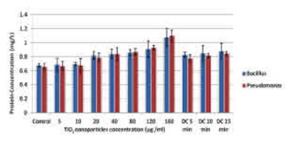
Figure 8: The eect of TiO2-NP concentrations (5- 160 ug/mL) and DC exposure (5, 10, and 15 min) on the protein concentration (mg/L) of B. cereus and P. aeruginosa biolms.
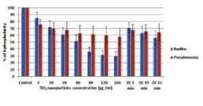
Figure 9: The eect of TiO2-NP concentrations (5-160 ug/mL) and DC exposure (5, 10, and 15 min) on the hydrophobicity percentage of B. cereus and P. aerugi- nosa biolms.
v Study of morphological changes in biolm before and after treatments
The use in this work of complementary microscopic means, such as SEM, enhanced visualization of morphological changes brought about by DC and NPs in the architectural properties of the mature preformed biolms (B. cereus and P. aeruginosa biolms), including cell surface, cell shape, cell distribution inside the EPS matrix, adhesion areas, and detachment. As depicted in Fig. 11, the mature biolm morphology and structure seemed to be unique for each biolm type examined. Both types of biolm appeared healthy, exhibiting normal rod cell shape with average dimensions: 1-0.2, 1.5-0.4 um length and 0.41-0.05, 0.2-0.05 um width for B. cereus and P. aeruginosa biolms, respectively (Figs. 11-A and 11-D). However, B. cereus cells were homogeneously distributed over the surface and fully wrapped in a dense, mucilaginous, stringy-like matrix of EPS (indicated by black arrows). On the other hand, P. aeruginosa cells were massive, compacted, smooth, aligned, tightly packed, and aggregated on an amorphous matrix. The SEM micrographs showed no signicant alterations in overall size or cell surface following the DC and NP treatments, although there was obvious destruction of the biolm structure. By applying DC with 9 V and 6 mA for 15 min, moderate damage was observed in B. cereus biolm.
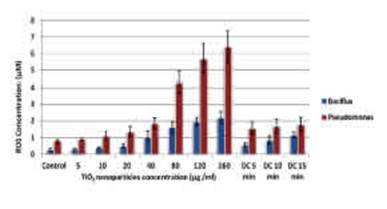
Figure 10: The eect of TiO2-NP concentrations (5-160 ug/mL) and DC exposure (5, 10, and 15 min) on the ROS concentration (uM) of B. cereus and P. aeruginosa biolms.
The majority of the cells retained the same normal shape with a lower number of cells individualized in less dense EPS matrix (Fig. 11-B). In addition, some cells were dramatically distorted, with clear furrows (indicated by red arrows); this implies disruption of the membranes of the bacterial cells and further leakage of cellular cytoplasmic uid, as recorded by Krishnamurthi et al., 2020 45. In contrast, relatively potent biolm destruction was displayed by P. aeruginosa biolm (Fig. 11-E). As shown, severe deformation was observed in the cell debris that still adhered to the slimy matrix. Our results are consistent with the earlier nding of Luo et al., 2005 46. Regarding TiO2-NPs treatment, Fig. 11-C illustrates aggregations of TiO2-NPs absorbed on the EPS-matrix of B. cereus biolm. Additionally, some cells appeared separate from each other, seemingly deformed by the presence of large furrows and small indentations (dashed yellow arrow). Similarly, Horst et al., 2010 47 recorded the agglomeration behavior of TiO2-NPs on biolm surface. Cell density markedly diminished with the damaged morphology (furrows and pits) of P. aeruginosa biolm exposed to TiO2-NPs (Fig. 11- F).

Figure 11: Scanning electron micrograph of B. cereus and P. aeruginosa biolms before and after DC and TiO2-NP exposure. (A) control biolm of B. cereus, (B) DC-treated B. cereus biolm, (C) TiO2-NPs- treated B. cereus biolm, (D) control biolm of P. aeruginosa, (E) DC-treated P. aeruginosa biolm, (F) TiO2-NPs-treated P. aeruginosa biolm. (Black arrows indicate EPS matrix, red arrows, furrows in damaged cells and yellow dashed arrows , indentations).
IV Discussion
Some studies have found that electrical current alone does not result in microbial death; however, other studies have reported some eect when applied to biolms. Poortinga et al. 48 reported electrical detachment of biolm formations from surgical implants, while Van der Borden et al. 49 demonstrated that DC of only 25{125 mA can stimulate detachment of staphylococcal strains from stainless steel, Moreover, Del Pozo et al. 14 recorded a decrease in the viability of S. aureus, S. epidermidis and P. aeruginosa biolms after prolonged exposure to a low-intensity electrical current of 20-2000 mA. On the other hand, Jass et al. 50 reported that electric currents of up to 20 mA/cm2 delivered for 12 hours did not prevent biolm formation or have any detrimental eect on an established biolm. Biolm formation can be reduced using lowintensity DC, but further investigation is needed to determine the appropriate dose and time of administration. The eectiveness of electric current in inhibition of growth and mortality is directly related to increasing microamperage 51. The high sensitivity of Gram-negative bacteria to electric current was conrmed by Davis et al. 52, who found that both E. coli and Salmonella typhimurium were inhibited and killed by low microamperage; they also reported that E. coli is more negatively sensitive to increasing current intensity than B. cereus. Our results support the theory that Gram-negative bacteria (P. aeruginosa) are more sensitive to low DC than Gram-positive bacteria (B. cereus), with a dierence of 9{11% for 5{15 min of exposure. This higher sensitivity is not only to DC but also to TiO2-NPs: The concentration of TiO2-NPs required to cause LD50 is lower for P. aeruginosa (63 -3 ug/mL) than for B. cereus (104-4 ug/mL). The development of biolm-related infections begins with adhesion of the microorganism to the biomaterial surface, mediated by Van derWaals forces, acid-base interactions, and electrostatic forces 53. The electrostatic force between bacteria and the biomaterial is generally repulsive, since almost all biomaterial surfaces are negatively charged, as are bacterial cells 54. It has been proposed that repulsive forces can be enhanced by the application of electric current, provoking surface detachment of bacterial biolms 55. When a biolm-covered steel slide was connected as the anode in an electrical circuit with a 6 V potential, biolm rapidly sloughed from the surface 17. DC alone had a lethal eect on both biolms in our study: 33% for B. cereus and 44% for P. aeruginosa after 15 min of DC exposure.
It has been proposed that the direct damagecaused to biolms by DC is by electroporation and/or production of ROS, as well as the generation of other toxic substances such as Chlorine 11. The eects of electrical currents on S. epidermidis biolms were interpreted by considering the electrolytic reactions occurring: it was hypothesized that an increase in pH near the anode leads to alkaline hydrolysis of the polysaccharide matrix of the biolm 17. Our results show that signi- cantly more ROS is produced in the DC-treated groups than in the control, for both biolms. Summarily, the substantial biocidal mechanism induced by DC is production of ROS (e.g., H2O2, chlorine molecules, etc.) as a result of electrolysis. This triggers enzyme oxidation and membrane puncturing, which leads to leakage of cytosolic constituents and a reduction in respiration rate 16. The DC had an indirect eect with prolonged time of exposure, through temperature and pH. This liberated, accelarated and oriented the charges/electron in the electrical eld toward negatively charged EPS. In turn, this impaired the biolm matrix stabilization, altered the surface charge, reduced hydrophobicity, perturbed bacterial membrane integrity, increased membrane permeability and increased ROS 56,57. All these destructive eects were emphasized in Figs. 7, 8, 9, 10, and 11.
In the current study, the ability of TiO2-NPs to disintegrate the biolm established by B. cereus and P. aeruginosa was exerted through this multidisruptive mechanism. It began mainly by disrupting and destabilizing the protective matrix (extracellular polymeric substances) containing mostly EPS and protein. This appears clearly in Figs. 7 and 8, and also in the reduction of hydrophobicity property as shown in Fig. 9, where a higher concentration of unattached EPS and protein content was eliminated when the concentration of TiO2- NPs was increased. Similarly, in the same context, hydrophobicity decreased with an increase in the treatment dose. The signicant relationship between cell surface hydrophobicity and biolm formation, which is closely linked to the EPS matrix secreted by the biolm 58, 59, is worthy of mention. In line with our results, Mu et al., (2021) 60 found that powerful treatment with antibiolm agents caused a decrease in S. epidermidis biolm attachment, and consequently decreased cell surface hydrophobicity via attenuation of EPS formation. Once TiO2-NPs have disturbed the EPS matrix, they enter the cells via porin channels and continue to damage them. Figures 10 and 11 support this. As TiO2-NPs were increased, the oxidative stress triggered by ROS increased. Additionally, the morphological changes caused by the TiO2-NPs, in the form of cell deformation and decreasing EPS content, were observed clearly by SEM (Fig. 11). All this evidence airms the antagonistic activity of TiO2-NPs. TiO2-NPs show antibacterial properties against Gram-positive and Gram-negative bacteria (7{8 nm), the latter being the more sensitive. This could be related to the fact that Gram-positive bacteria have a thicker layer of peptidoglycan (20{80 nm) than Gram-negative bacteria, which facilitates the absorption of reactive radicals, thereby preventing cell damage from radical attack 31, 34.
TiO2-NPs can reduce the adhesion of bacteria and inhibit biolms. Exposure to TiO2-NPs leads to the destruction of bacteria inside the biolm, primarily due to the generation of ROS and lipid oxidation on the cell wall membrane 61. It has been shown that TiO2-NPs are eective against biolms of MRSA 62. TiO2-NPs could control the growth and biolm formation of S. mitis ATCC 6249 and Ora-20, and can be used in oral hygiene. TiO2- NPs have a low impact on P. aeruginosa biolms at 31.25 ug/mL concentration and disrupt previously established biolms in the microtiter plate 63. In the presence of TiO2-NPs, the biolm formation of E. coli and B. subtilis was reduced by 40-50% respectively 64. However, TiO2-NPs did not show signicant bactericidal properties against certain types of drug-resistant bacteria which have a remarkable ability to withstand ROS membrane damage through over-expression of protective components and membrane repair elements 65. Our results indicate that there is a signicant increase in ROS production in groups exposed to 20 ug/mL or more, and this eect increases as TiO2-NP concentration increases.
Hydrophobic interactions in bacteria are one of the most important mechanisms for microbial attachment and aggregation, which are strongly associated with the protein secondary structures on the cell surfaces. The changes in the protein secondary structures on bacterial surfaces aected these hydrophobic interactions, which reduced the bacterial attachment ability 66. The dynamic response of a B. subtilis biolm to temporary exposure to TiO2-NPs caused the dispersal of biolm and bacteria after several hours of exposure, indicating that the changes in the cell/EPS surface structure and the decreased adhesive ability drove the biolm dispersal 67. Our current results show that the hydrophobicity of both biolms is reduced by using either DC or TiO2-NPs of concentration 10 g/mL or more. In a previous study, TiO2-NPs showed lower inhibitory and biolm concentration against S. mu- tans and S. sanguinis than NPs containing Ag NPs, Fe3O4 NPs, antibiotics, and chlorhexidine 68. TiO2-NPs also exhibit marked antimicrobial and antibiolm activity against ATCC 6249 and Ora-20 and hence can control their growth and biolm formation in the oral cavity even at a concentration as low as 50 ug/ml, due to disruption of the cell wall and oxidative stress 69, which are recorded also in the current study. Generally, although the EPS-matrix represents the robust skeleton that protects the biolm cells from stress, the eradication and detachment capacity of both DC and NPs were evident throughout the current study. This could be attributed to alterations in the physical-chemical characteristics of both biolm and adherent surfaces (i.e., polymeric properties, hydrophobicity/hydrophilicity, charge, roughness, and surface free energies) induced by both treatments, which ultimately destabilized adhesion of the preformed biolm to the surface 70, 71. Moreover, the involvement of water channels in the core structure of the biolm, which allow mainly the transportation of nutrients, could permit the diusion of toxic substances that generate ROS, which unambiguously cause cell damage 71{73. This assumption was conrmed simultaneously via SEM (the presence of pores, pits, furrows, and cell deformation) and ROS results. Interestingly, the absence of full inhibition and eradication of both biolms by DC and NPs could be explained by the higher resistance of mature biolm cells during the stationary phase, as reported by Rodrigues et al. 74. Arguably, any antibiolm treatment will exhibit higher potency when applied during the evolution of a biolm than when applied to a mature preformed one. Microbial cells that are freeoating or at the early stages of colonization seem to be vulnerable and susceptible to any treatment, especially before formation of the EPS-barrier and the evolution of quorum sensing signals between colonized cells 75.
V Conclusions
Our results indicate that both DC and TiO2-NPs have a lethal eect on Gram-Positive and Gram- Negative bacterial biolms. Gram-Negative bacterial biolms are more sensitive to both DC and TiO2-NPs than the Gram-Positive ones. Applying DC of 16.2{48.6 J aects bacterial biolms in a similar way of using TiO2-NPs of 20-40 ug/mL concentration. TiO2-NP concentrations higher than 40 ug/mL produce a signicantly greater lethal effect than the DC conditions used on both biolms. The lethal eect on biolms was veried by EPS, protein content and cell surface hydrophobicity assessment, as well as scanning electron microscopy visualization. The mechanism of action was correlated with the ROS produced.

