


Services on Demand
Journal

Article
 English (pdf)
 Article in xml format
 Article references
 Send this article by e-mail
Indicators
-
 Cited by SciELO
Related links
-
 Similars in
SciELO
Similars in
SciELO
Share

 Permalink
Acta Odontológica Latinoamericana
On-line version ISSN 1852-4834
Acta odontol. latinoam. vol.24 no.3 Buenos Aires Dec. 2011
ARTÍCULOS ORIGINALES
Cephalometric norms from posteroanterior Ricketts’ cephalograms from Hispanic Americans Peruvian non adult patients
Iván E. Pérez1, Allison K. Chávez2, Darío Ponce3
1 Oral Radiology Department. Dentofacial disarmonies research center.
2 Department of Endodontics. Cayetano Heredia Peruvian University Dental School of Dentistry.
3 Statistical Department. Dentofacial disarmonies research center.
CORRESPONDENCE Ivan Perez Lip CIDDENT S.A. iepl76@yahoo.com Av. Arequipa 4252 Oficina 1 Lima 18, Lima – Peru
ABSTRACT
Aim: The purpose of the present study was to describe the posteroanterior cephalometric norm values from Hispanic Americans Peruvian non adults patients between years 2009 to 2010, identify possible differences between sexes and compare our results with similar studies in the literature. Material and methods: Data from posteroanterior cephalograms from 318 patients (177 females and 141 males) between 9 and 18 years old were collected from our database; mean and standard deviation were calculated for each gender and age group. Results: Independent samples T-test found statistically significant differences between males and females results in the intermolar width, right molar to maxillae distance, nasal width, nasal height, maxillary width, mandibular width and facial width. Conclusions: statistically differences between sexes were found in seven from twelve transversal measurements. The norm values found in this study are similar to those reported by Ricketts’.
Keywords: Cephalometry; Reference values; Radiography; Dental; Orthodontics; Population characteristics.
RESUMEN
Valores promedio de la cefalometría posteroanterior de Ricketts en pacientes no adultos Peruanos Hispano Americanos
El propósito del presente estudio fue de establecer valores promedio de las medidas transversales del análisis posteroanterior de Ricketts en pacientes Peruanos Hispano Americanos no adultos que asistieron a nuestro centro de radiología entre los años 2009 y 2010, identificar posibles diferencias entre géneros y contrastar nuestros hallazgos con estudios similares en la literatura. Materiales y método: Se recolectaron los valores de 12 medidas transversales 318 análisis de cefalometría posteroanterior de Ricketts (177 mujeres y 141 hombres) de nuestra base de datos; para cada medida se calculó el promedio y la desviación estándar de cada género y cada grupo etáreo. Resultados: La prueba estadística T encontró diferencias estadísticamente significativas entre las medidas de los géneros masculino y femenino en el ancho intermolar, distancia del molar derecho al maxilar, ancho nasal, altura nasal, ancho maxilar, ancho mandibular y ancho facial. Conclusiones: Se encontraron diferencias estadísticamente significativas en las medidas ancho interpolar (IM), distancia del molar derecho al maxilar (RMMD), ancho nasal (NC-CN), altura nasal (NH), ancho maxilar (JL-JR), ancho mandibular (AG-GA) y ancho facial (ZA-AZ).Los valores encontrados en la población estudiada son similares a los valores promedio reportados por Ricketts.
Palabras clave: Cefalometría; Valores de referencia; Radiografía; Dental; Ortodoncia; Características de población.
INTRODUCTION
Cephalometric norms have been developed for the different ethnic and racial groups and it has been concluded that there are significant differences between them. Different racial groups must be treated according to their own characteristics1. Posteroanterior (PA) cephalometric analysis is used to: evaluate the vertical, transverse and sagittal dimensions1, diagnose and quantify facial asymmetries and skeletal abnormalities2, assess the transverse changes induced by maxillary expansions3, and diagnose functional, dentoalveolar, and/or facial asymmetries4. The relationship between the widths of the maxillary and mandibular skeletal bases is the most critical information from the posteroanterior cephalometric analysis1. Most of the normative data have been based on lateral cephalometric radiographs and provides information on sagittal aspects of dentofacial structures5. Facial asymmetries and the oronasal area development can be assessed with the transverse analysis of PA cephalometric radiographs5.
The use of PA cephalometry is not standardized like lateral cephalometry2. Researchers have been reluctant to use PA cephalometry for a variety of reasons such as: difficulties in reproducing head posture and landmark identification due superimposition or poor radiographic technique3,4. Some of those difficulties could be overcome by careful attention to radiographic technique and selection of skeletal and dental landmarks with acceptable reliability3. Transverse measurements or widths from PA cephalograms are least affected by positional errors3. Cephalometric analysis errors are classified in: radiographic projection, landmark identification, tracing and measurement errors4. Cephalometric points located on a sharp curve or at the intersection of two curves are, generally, easier to identify than those located on a flat or broad curve4, and, cephalometric points located in high contrast areas are easier to identify than the low contrast ones4. In the PA cephalometry, vertical and transverse measurements of dentofacial structures are taken relative to reference lines and the asymmetry are calculated by comparing the measurements of corresponding structures from the left and right sides8. The midline structures can also be used to asses the asymmetry as deviation toward the right or left side from the chosen line of reference8. The Ricketts’ PA cephalometric analysis seems to be the most widely used because it provides normative values for different ages.1,9
The aim of this study was to describe the cephalometric mean values from Ricketts’ PA cephalograms from Hispanic Americans Peruvian non adult patients, identify possible differences between both genres and compare our results with similar design studies in the scientific literature.
MATERIAL AND METHOD
Data was collected from PA cephalograms of Hispanic Americans patients between 9 to 18 years old from Lima – Peru, who attended our radiology center (CIDDENT) for radiographic assessment prior to orthodonctis between years 2009 - 2010. PA radiographs were taken in an Odontorama PC 100 panoramic machine (Trophy - France) in maximum intercuspation and the Frankfurt plane parallel to the floor. The cephalometric radiographs were traced on acetate cephalometric tracing sheet (GAC - cephalometric tracing paper) by a trained professional not associated to the present study. From the 429 PA cephalograms selected, 111 were excluded because they met the following exclusion criteria: data absence, error in data entry, craniofacial syndromes, cleft palate (any type), and absence of fist molars, lower canines, or incisors. As a result, 318 PA cephalograms were available to perform this study. We collected data from 12 skeletal and dental linear measurements from Ricketts’ PA cephalometric analysis in millimeters (Tables 1, 2; Figures 1, 2).
Table 1: Posteroanterior cephalometric landmarks.
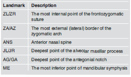
Table 2: Posteroanterior cephalometric reference planes.
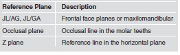
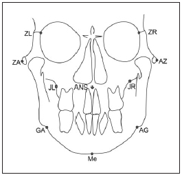
Fig. 1: Posteroanterior cephalometric landmarks.
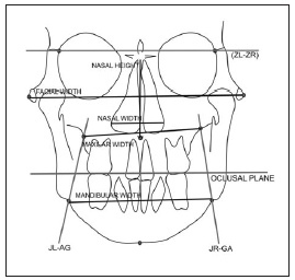
Fig. 2: Posteroanterior cephalometric reference planes.
Data was grouped in MS Office Excel 97 and statistical analyses were performed on IBM SPSS Statistics 15. For the 12 selected linear measurements of Ricketts’ PA analysis mean, standard deviation and variance ware calculated. Ten PA radiographs were select randomly and were traced twice in a one week interval. We used the method of error formula found in the study of Ishiguro et al6, the result was less than 1 mm.
RESULTS
The data was comprised of PA cephalometric analysis of 318 PA cephalograms, 177 females and 141 males between 9 and 18 years of age. The mean age value was 12.60 +/- 2.18 years old; the female mean age value was 12.28 +/- 2.17 years old and the male mean age value was 12.99 +/- 2.14 years old. (Tables 3-8)
Table 3: Population age and gender distribution.
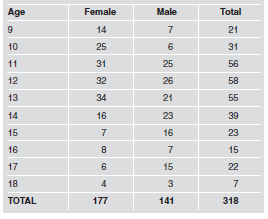
Table 4: Descriptive statistics of posteroanterior cephalometric measurements (in millimeters) for non adults Hispano American Peruvians.
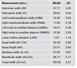
Table 5: Descriptive statistics of posteroanterior cephalometric measurements (in millimeters) for a non adult Peruvian women.
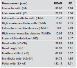
Table 6: Descriptive statistics of posteroanterior cephalometric measurements (in millimeters) for a non adult Peruvian men.
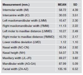
Table 7: Descriptive statistics of posteroanterior cephalometric measurements (in millimeters) for a non adult Peruvian by age.
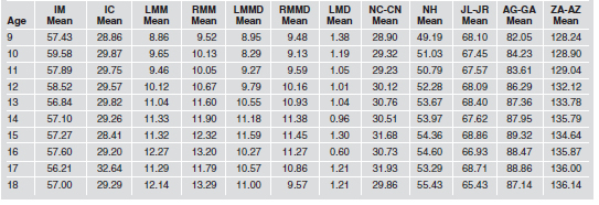
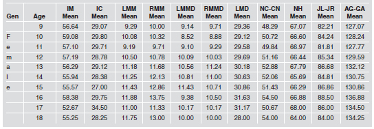
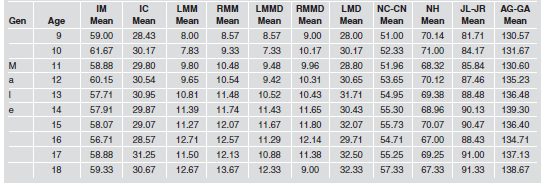
Table 8: Descriptive statistics of posteroanterior cephalometric measurements (in millimeters) for a non adult Peruvian by gender and age.
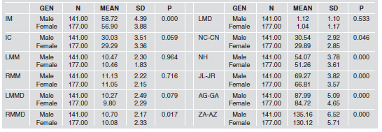
The independent samples t-test was used to compare differences between sexes, the results found statistically significant differences in the intermolar width (P <0.000), right molar to maxillae distance (P <0.017), nasal width (p <0.046), nasal height (p <0.000), maxilar width (p <0.000), mandibular width (p <0.000) and facial width (P <0.000). (Table 9)
Table 9: T test results of posteroanterior cephalometric measurements (in millimeters) between men and women.
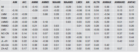
To study the correlation between each of the 12 PA measurements, Pearson correlation coefficients were calculated. The highest coefficient value was found between left and right maxillomandibular width (r = 0.68), the lowest correlation was found between maxilar width and right maxilomandibular width (r =- 0.36). (Table 10)
Table 10: Pearson’s correlation coefficient of posteroanterior cephalometric measurements.
DISCUSSION
The importance of the transverse dimension becomes apparent when the potential and limits of certain treatment options, such as palatal expansion, have to be explored, or when deciding between the extraction or non extraction in borderline cases9. Transverse and vertical distances demonstrated a progressive increase between 10 and 14 years in both genres, with vertical growth greater than the transverse facial growth5. Facial growth finishes first in width, then in length and finally in height5, 7. Publications concerning normative data related to PA cephalometry in different non adult populations were made by Athanasiou12 (Austria), Moyers (USA), Ricketts (USA), Cortella7 (USA); studies in adult population were conducted by Wey (China), Uysal and Zafer1 (Turkey) and Altaki13 (Palestine). Intermolar width (IM) mean value found in this study was 57.71± 4.2mm, the female population mean value was 56.9± 3.88mm and the male population mean value was 58.72± 4.39mm. Our results are similar to those reported by Ricketts, Zafer and Uysal1 in the Turkish adult population and Altaki in the Palestinian adult population13. The mandibular canine width (IC) mean value found in this study was 26.92± 3.44mm, the female population mean value was 26.29± 3.36mm and the male mean value was 30.3± 3.51mm. Our results are similar to Ricketts norm value and to those reported, in the general population and female population, by Uyzal and Zafer1 in the Turkish population, the male mean value found in this study was higher when compared to Uyzal and Zafer1 findings in the Turkish male adult population.
Maxilomandibular width (LMM and RMM) mean value found in this study for the left side was 10.46± 2.05mm; the female population mean value was 10.46± 1.83mm and the male population mean value was 10.46± 2.30mm. The right side the mean value was 11.08± 2.18mm, the female population was 11.05± 2.15mm and the male population mean value was 11.13± 2.22mm. Our results are similar to those reported by Ricketts and lower to those reported by Uysal and Zafer1 in the Turkish male adult population. The molar to maxillae distance (LMM) mean value found in this study for the left side was 10.01±2.39mm; the female population mean value was 9.8± 2.29mm and the male population mean value was 27.10±2.49mm. The right side mean value was 10.36±2.28mm; the female population mean value was 10.08± 2.33mm and the male population mean value was 10.7±2.17mm. Our results are higher to those reported by Ricketts and similar to those reported by Uysal and Zafer1 in the Turkish adult population. The nasal width (NC-CN) mean value found in this study was 30.18± 2.89mm; the female population mean value was 29.89± 2.85mm and the male population mean value was 30.54± 2.92mm. Our results are lower than those reported by Uyzal and Zafer1 in the Turkish adult population, Altaki13 in the Palestinian adult population and similar to those reported by Wei14 in the Chinese adult population. In Ricketts’ PA analysis, the nasal width norm value varies between 25 to 31.3mm among 9 to 18 years old, our results found range values between 28 to 31mm, which are similar to those reported by Ricketts. The maxillary width (JL-JR) mean value found in this study was 67.9± 3.87mm; the female population mean value was 66.81± 3.57mm and the male population mean value was 69.27± 3.82mm. Our results are similar to those reported by Uyzal and Zafer1 in the Turkish adult population and Altaki13 in the Palestinian adult population; our results for the female population are higher than those reported by Uyzal and Zafer1. In the Ricketts’ PA analysis, the norm value varies between 62 to 67.4mm among 9 to 18 years old, our results found range values between 65 to 68mm, similar to those reported by Ricketts.
The mandibular width (AG-GA) mean value found in this study was 86.17± 5.11mm; the female population mean value was 84.72± 4.65mm and the male population mean value was 87.99± 5.09mm. Our results are lower than those reported by Uysal and Zafer1 for the Turkish adult population and similar than those reported by Altaki13 for the Palestinian adult population, Cortella7 and Ricketts. Facial width (ZA-AZ) mean value found in this study was 132.36± 6.57mm; the female population norm value was 130.12± 5.71mm and the male population value was 135.16± 6.52mm. Our results are higher than those reported by Wei14 in the Chinese adult population, lower than those reported by Uysal and Zafer1 in the Turkish adult population and similar to those reported by Altaki13 in the Palestinian adult population and the Ricketts’ PA analysis. Statistically significant differences between male and female genres was found in the intermolar width (IM), right molar to maxillae distance (RMMD), nasal width (NC-CN), nasal height (NH), maxillary width (JL-JR), mandibular width (AGGA), facial width (AZ-ZA). Uysal and Zafer1, Wei14, Yavuz5 and Altaki13 found differences between genres in facial width, nasal width and maxillary width respectively. Wei14 and Uysal5 found differences in the intercanine width. Uysal and Zafer1 and Altaki13 found differences in the intermolar width and mandibular width. Our study population consisted of cephalograms from the Hispanic American Peruvians ethnic group; ethnic line studies can be developed to collect data from two previous generations to make comparisons and establish so microevolutionary changes. The real cephalometric norm values for our ethnic group can be found with similar design studies in the three geographical regions of our country. Differences between male and female genres found in our study can be supplemented with other cephalometric measurements for a better description of secondary sex characteristics.
CONCLUSIONS
Differences between sexes were demonstrated in seven from twelve transversal measurements, especially in the internal structural field of the Ricketts analysis. The mean values found in this study are similar to those reported by Ricketts, with exception bilateral molar to maxillae distance (MMD); so it is advisable to use the Rickett´s PA cephalometric norm values . Differences between populations were found by comparing studies of similar design.
REFERENCES
1. Uysal T, Zafer S. Posterior cephalometric norms in Turkish adults. Am J Orthod Dentofac Orthop. 2005;127:324-332. [ Links ]
2. Kirjavainen M, Kirjavainen T. Maxillary Expansion in Class II Correction with Orthopedic Cervical Headgear. A Posteroanterior Cephalometric Study. Angle Orthod. 2003;73: 281-285. [ Links ]
3. Cross D, McDonald JP. Effect of rapid maxillary expansion on skeletal, dental, and nasal structures: a postero-anterior cephalometric study. Eur J Orthod. 2000;22:519-528. [ Links ]
4. Leonardi R, Annunziata A, Caltabiano M. Landmark Identification Error in Posteroanterior Cephalometric Radiography. Angle Orthod. 2008;78:761-766. [ Links ]
5. Yavuz I, Ikbal A, Baydas B, Ceylan I. Longitudinal Posteroanterior Changes in Transverse and Vertical Craniofacial Structures Between 10 and 14 Years of Age. Angle Orthod. 2004;74:624-629. [ Links ]
6. Ishiguro K, et al. A Longitudinal Study of Morphological Craniofacial Patterns via P-A X-Ray Headfilms in Cleft Patients from Birth to Six Years of Age. Cleft Palate Journal. 1976;13:104-126. [ Links ]
7. Cortella S, Shofer FS, Ghafari J. Transverse development of the jaws: Norms for the posteroanterior cephalometric analysis. Am J Orthod Dentofac Orthop. 1997;112:519-522. [ Links ]
8. Trpkova B et al. Assessment of facial asymmetries from posteroanterior cephalograms: Validity of reference lines. Am J Orthod Dentofac Orthop. 2003;123:512-520. [ Links ]
9. Lux CJ, Conradt C, Burden D, Komposch G. Transverse development or the craneofacial skeleton and dentition between 7 and 15 years of age – a longitudinal posteroanterior cephalometric study. Eur J Orthod. 2004;26: 31-42.
10. Snodell SF, Nanda RS, Currier GF. A longitudinal cephalometric study of transverse and vertical craniofacial growth. Am J Orthod Dentofacial Orthop. 1993;104:471-483. [ Links ]
11. Giuca MR, et al. Correlations between transversal discrepancies of the upper maxilla and oral breathing. Eur J Paediatric Dent. 2009;10:23-29. [ Links ]
12. Athanasiou AE, Droschl H, Bosch C. Data and patterns of transverse dentofacial structure of 6 to 15 year olf children: A posteroanterior cephalometric study. Am J Orthod Dentofac Orthop. 1992;101:465-471. [ Links ]
13. Al Taki A. Dentofacial Transverse Dimensions in Palestinian Adults. Smile dental Journal [internet]. 2009;4:6-10. http://www.smiledentaljournal.com/index.php?option=com_content&view=article&id=114&Itemid=110. [ Links ]
14. Wei SHY. Craniofacial width dimensions. Angle Orthod. 1970; 40: 14-147. [ Links ]

