


 English (pdf)
 Article in xml format
 Article references
 Send this article by e-mail
 Cited by SciELO
  Similars in
SciELO
Similars in
SciELO 
 Permalink
INTRODUCTION
Universal dental adhesives were introduced as versatile multifunctional systems with fewer application steps, compatible with all treatment modalities for mineralized dental tissues1. Universal adhesives, also known as multimode or multipurpose adhesives, have been used in clinical practice since 2011. They can be applied to enamel and dentin as self-conditioning adhesives, and to enamel as etch-rinse adhesives, a technique known as “selective enamel etching”. The acid monomers in their composition act as primers for non-dental substrates (alloys and polycrystalline ceramics), making universal adhesives suitable for application with different bonding strategies2,3. These adhesives have been recently called eighth-generation adhesives, based on the historical perspective of dental adhesives4. Universal adhesives contain mixed monomers with slight to moderate acidity in reduced concentrations, conventional dimethacrylate cross-linkers, non-acid emulsifying monomers, catalyzer for light and dual curing and an adequate selection of solvents to improve monomer spreading and substrate infiltration capacity2,5.
An adhesive that may be used in different procedures enables the dentist to choose the technique that best suits the clinical case, thereby optimizing the final restorative result. For instance, when a restoration requires a strong bond to the enamel, or in case of sclerotic dentin, it may be advisable to perform previous etching. The etching stage can be graded according to the time for which the phosphoric acid gel is applied before rinsing. On the other hand, it may be preferable to use the self-conditioning technique when dealing with difficult surgical access, limited time, or non-collaborative very young patients6. Even though it has been scientifically documented that the etching technique using phosphoric acid enhances one-step self-conditioning bonding to enamel, it is advisable to take more care with the etching procedure using additional phosphoric acid on dentin7. This is probably the main reason why most universal adhesive contain 10-methacryloxydecyl dihydrogen phosphate (10-MDP), a functional group of phosphoric acid, as their main adhesive monomer which produces a limited decalcification effect on the dentin surface. This procedure is considered the most reliable treatment for dentin, since the universal adhesives containing MDP have slight acidity and the capacity to interact chemically with hydroxyapatite crystals and enable stable salt formation of calcium phosphate and calcium carboxylate insoluble in water8,9.
This new philosophy of versatile bonding encourages the use of the simplest option for each strategy, i.e., one-step self-conditioning or two-step etch and rinse, in order to bond direct or indirect restorations to enamel and dentin10. The universal etch and rinse (E&R) bonding mode involves a phosphoric acid etching step followed by a thorough water rinsing phase prior to application of a primer/adhesive resin combination. Monomers diffuse into the micro-etch pits created on the enamel to form microtags and macrotags, and into the exposed collagen fibril network at dentin to form a 3-5 pm hybrid layer. While the E&R bonding mode is undoubtedly the best bonding strategy to enamel, the resultant thick and HAp-free hybrid layer formed on the dentin is highly sensitive to degradation over time. The universal self-etch (SE) bonding mode involves the use of monomers with an acidic functional group that in principle simultaneously etches and infiltrates dentin down to a depth of about 1 pm. In general, the SE bonding mode underperforms the E&R bonding mode on enamel, by which enamel remains to be selectively etched with phosphoric acid. SE bonding nevertheless possesses chemical bonding potential as an additional benefit to achieve durable bonding4. However, universal bonding systems may show the deficiencies of their predecessors, i.e., one-step systems11, so their bonding performance to dentin should be evaluated using different variables that modify in-vitro bond strength12. In this context, the purpose of the present study was to use shear bond strength and laser microscopy tests to determine the bonding characteristics of universal adhesives using total enamel and dentin etching treatments, adamantine selective etching and self-conditioning on the pulp wall of deep cavity preparations.
MATERIALS AND METHOD
This study was approved by the Academic Committee for Health Research (Comité Académico de Investigaciones en Salud) of the School of Dentistry, National University of Cordoba (CIEIS-ODO-CASI 48 I).
Cavity preparation
Single occlusal cavities 4 mm deep and maximum width were carved in 36 third molars which had been extracted for orthodontic reasons. Cavity depth standardization was checked using a periodontal millimeter probe, CP-11, Hu-Friedy, Illinois, United States. The molars were divided into nine groups according to dentin substrate treatment (total etching, selective adamantine etching or self-conditioning), and the universal adhesive applied. Group 1: Total-etch+Monobond 7 self-etch adhesive (Densell, Buenos Aires, Argentina). Group 2: Total-etch+One coat 7 universal adhesive (Coltene, Altstatten, Switzerland). Group 3: Total-etch+Single bond universal adhesive (3M ESPE, Neuss, Germany). Group 4: Adamantine etching+Monobond 7 self-etch adhesive (Densell, Buenos Aires, Argentina). Group 5: Adamantine etching+One coat 7 universal adhesive (Coltene, Altstatten, Switzerland). Group 6: Adamantine etching+Single bond universal adhesive (3M ESPE, Neuss, Germany). Group 7: Monobond 7 self-etch adhesive (Densell, Buenos Aires, Argentina). Group 8: One coat 7 universal adhesive (Coltene, Altstatten, Switzerland). Group 9: Single bond universal adhesive (3M ESPE, Neuss, Germany). In groups 1, 2 and 3, total-etch was performed on enamel and dentin using 35% phosphoric acid (Densell, Buenos Aires, Argentina) for 10 s, followed by spray washing and drying for 5 s. Finally, universal bonding was applied according to the manufacturer’s instructions. In groups 4, 5 and 6, enamel was etched using 35% phosphoric acid (Densell, Buenos Aires, Argentina) for 10 s. In groups 7, 8 and 9, universal adhesives were applied directly on-to the enamel and dentin as indicated by the manufacturer.
All the cavities were filled with submicron-hybrid composite resin Brilliant EverGlow (Coltene, Altstatten, Switzerland), by means of oblique incremental technique, and each layer was polymerized for 20 s with a LED unit (Optilux LED, Coltene, Altstatten, Switzerland).
Shear bond strength
Three specimens from each group (27 altogether) were first sectioned longitudinally in lingual-buccal direction into sections 2.5 mm and 3 mm thick on average; then transversally, using a hard tissue microtome Isomet (Buehler Co., Illinois, United States) at 300 rpm and 50 grams pressure, under continuous water cooling. Two test specimens averaging 2.6 mm wide by 2.6 mm high were thus formed, which included dentin pulp wall, universal adhesive system and composite resin. Thus, each group consisted of six test specimens (n=6). These samples were stored at 37 °C for 24 h in a 100% humidity atmosphere.
Shear bond strength tests were performed using a universal testing machine with a crosshead speed of 0.5 mm per minute. The sections were fixed with a dental press and placed on the plate of the device such that the composite resin/dentin pulp wall union remained next to the edge. The beveled edge of the shear was placed at 0.5 mm from the material/substrate joint for cutting. A standard 0.5 mm thick layer was used to corroborate the separation. Analysis of variance was applied to determine the efficacy of the substrate treatment with each universal adhesive used.
Laser confocal microscopy study
One specimen from each group (9 altogether) was sectioned longitudinally using a hard tissue microtome Isomet (Buehler Co., Illinois, United States) at 300 rpm and 50 g pressure under continuous water cooling, to obtain test specimens 1.5 mm to 2 mm thick for each group (n=3 for each group). The sections were polished using a metallographic polisher (Praxis, Buenos Aires, Argentina) with tapered granulometry disks and felt cloths, washed with ultrasound and stored in a stove at 37 °C for 24 h in a 100% humidity atmosphere. The sections were observed with laser confocal microscopy, Lext 3D Measuring Laser Microscope OLS4000 (Olympus, Japan). The thicknesses of the adhesive layer on the deep pulp walls were measured using the same software. Ten measurements were performed by microphotography at regular intervals on 50 pm long paths ( Fig. 1 ). The data were analyzed using analysis of variance. Statistical significance was set at 0.05 for all tests.
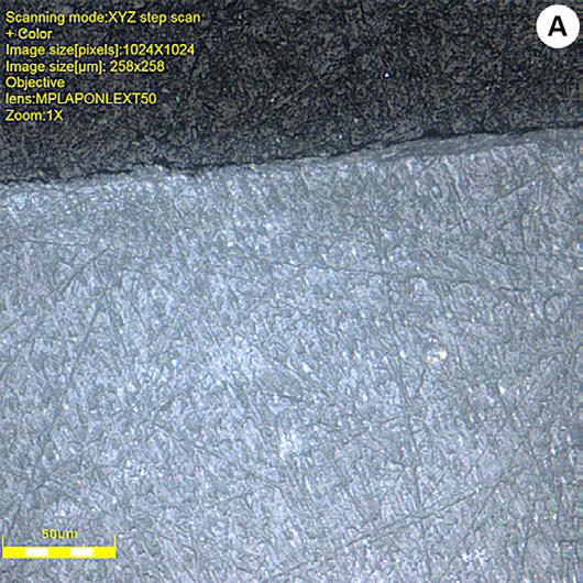
Fig. 1 B and C show profile sections that were measured A) Group 1: Simultaneous etching technique of enamel and dentin and application of Universal Adhesive Monobond 7 self-etch. Mean layer thickness between the pulp wall and the restoration material was 8.71±4.93 μm. B) Group 5: Application of One Coat Universal Adhesive by means of selective enamel etching. Mean layer thickness was 5.49±1.70 μm, and C) Application of Single Bond Universal Adhesive through self-etch mode. Mean layer thickness was 6.27±3.01 μm.
RESULTS
Shear bond strength
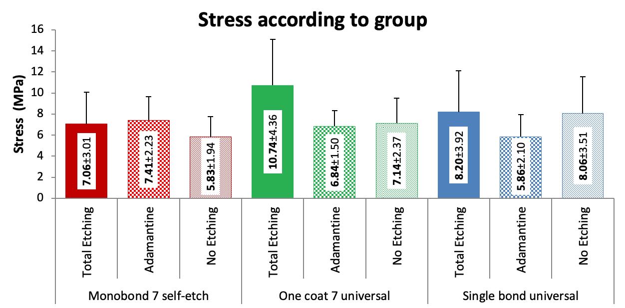
Considering each adhesive with its respective treatment, shear bond strength was significantly higher (p=0.024) for One coat 7 universal using total-etch procedure than for any of the other three treatments performed on the dental substrate. Monobond 7 self-etch had the highest values with selective enamel etching, with no significant difference (p=0.384). Single bond universal with total-etch protocol had higher valúes than the other treatments, with no statistically significant difference (p=0.299) ( Fig. 2 ).
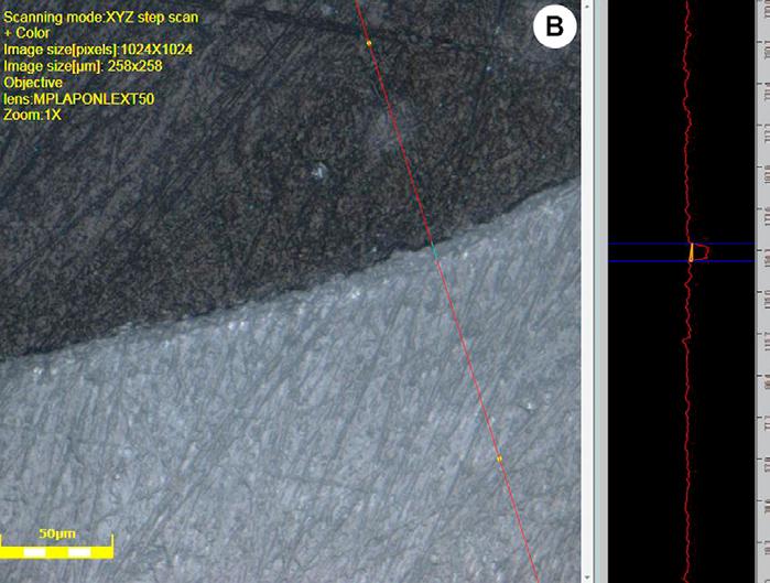
Fig. 2 Shear stress according to adhesive and technique used. Mean and standard deviation values expressed in MPa.
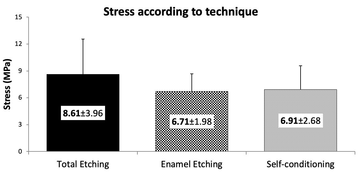
In general, bond strength for the total-etch treatment (8.61±3.96) was significantly greater than for the other treatments: selective enamel etching (6.71±1.98) and self-conditioning (6.91±2.68); p=0.049 ( Fig. 3 ).
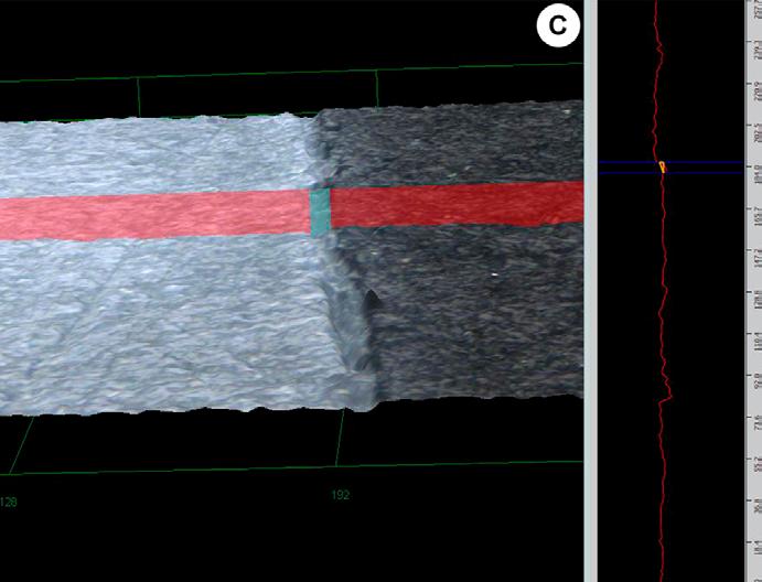
Fig. 3 Shear stress according to technique. Mean and standard deviation values expressed in MPa. Fig.
There was no statistically significant difference (p=0.205) among the different universal adhesives used. One coat 7 universal (8.33±3.45) mean values were higher than those of the other universal adhesives, Single bond universal (7.38±3.33) and Monobond 7 self-etch (6.75±2.47) ( Fig. 4 ).
Laser confocal microscopy analysis: Adhesive layer thickness
Adhesive layer thicknesses were greatest for the total-etch treatment group, followed by self-conditioning, and lowest for the selective enamel etching treatment group. There was no significant difference among the different adhesive techniques ( Fig. 5 ).
DISCUSSION
The present study found that certain in-vitro conditions such as deep pulp wall and different universal adhesive bonding approaches determined low bonding values expressed in MPa in the shear bond strength tests when compared to those obtained, for example, for lateral, median or superficial walls13. This means that cavity depth and wall type affect the results of the laboratory tests, regardless of the bonding approach chosen. We agree that it is more difficult to achieve bonding to the dentin closer to the pulp chamber roof than to the superficial dentin4. Adhesion to dentin remains a challenge, mainly because dentin has a more organic composition than enamel, and its wet, organic nature makes bonding difficult13. When dentin is etched, the acid demineralizes the intertubular dentin, resulting in the exposure of the superficial collagen network14.
The network is infiltrated with the adhesive resin, leading to the formation of a hybrid layer which is responsible for the bond between the resin and the dentinal tissues15. To ensure ideal bonding conditions, the dentin demineralized with acid must be kept wet to prevent the collagen fibrils from collapsing, though not too wet, because over-wetting prevents resin impregnation of the collagen fibrils16. Smear layer removal with single-step etching before the dentin adhesive is applied (etch-rinse technique), or its modification with a self-etch monomer (self-conditioning technique) is crucial to form a hybrid layer in order to ensure an effective bond between the adhesive resin and the dentin17.
The present study found higher valúes for total-etch treatments in deep dentin than for the other two methods (selective etching and self-conditioning). However, during the total conditioning step, phosphoric acid eliminates the smear layer while demineralizing the dentin to a depth of 3 to 5 mm, thus exposing a collagen fibril scaffold deprived of hydroxyapatite18,19. The highly mineralized peritubular dentin dissolves almost completely and the dentin tubules widen. However, the literature reports that no statistically significant difference was recorded in the dentin regarding bonding efficacy when self-etch or etch-rinse approaches were used2. It has been suggested that the bonding techniques requiring smear layer removal are associated with greater postoperative sensitivity than systems that leave the smear layer in situ20. Even though it has been demonstrated that the bonding procedure may cause transitory pulp inflammation, especially in the deep cavities, it is likely that continuous bacterial irritation due to microgaps and microfiltration may cause damage to the pulp and postoperative pain21. Shear bond strength is a valid versatile method for assessing bonding effectiveness to dental substrates in laboratory tests22. The formation of smear layer in vitro has also been shown in laboratory tests. Chowdhury et al. assessed the effects of the smear layers formed by abrasives having similar coarseness to the fine-grit diamond stone. They established a model for dentin bonding tests, yielding clinically relevant significant results. They also performed micro-tensile bond strength tests (pTBS) of currently available universal adhesives and of a two-step self-etch adhesive. The presence of smear layer formed under these conditions had no significant effect on the resin-dentin bond strength of the adhesives tested. Moreover, the performance of the bond of the universal adhesives to the dentin may be improved duplicating its application time. The elimination or modification of the smear layers covering the dentin is critical to allow penetration of the adhesive molecules and to ensure a strong bond between the resin and the dentin23. Universal adhesives benefit from phosphoric acid etching because bond strengths increase, mainly on the enamel surface. The authors compared in vitro shear strength of four universal adhesives on enamel and dentin with and without additional phosphoric acid etching, finding mean bond strength values to enamel ranging from 13.4 and 21.9 MPa in the self-etch mode. When the etch-rinse protocol was used, the mean bond strength was over 30 MPa. Regarding the dentin, the significant differences in the self-etch mode depended on each adhesive used24. Stape et al. claim that selective dentine etching for 3 s improved the interaction depth of the tested universal adhesive without overexposing the demineralized collagen or reducing Ca availability at the bonded interface. Nevertheless, the universal adhesives used in the self-etch mode produce superior long-term dentin bonding compared to the etch and rinse mode. Selective etching for 3 s with conventionally used H3PO4 improves dentin bonding effectiveness; nonetheless, longer etching times should be strictly avoided25. With regard to the self-conditioning protocol, Tsuj imoto et al. compared universal adhesives in self-conditioning mode and two-step self-conditioning adhesives by means of initial shear bond strength tests and shear-fatigue strength test, at the level of the resin composite/adhesive bond to dentin. Their results encourage the continued use ofthe two-step self-etch adhesive over some universal adhesives but suggest that changes to the composition of universal adhesives may lead to dentin bond fatigue durability similar to that of two-step self-etch adhesives26. Daneshkazemi et al. reported mean values in MPa between 35.74 and 18.09 in superficial dentin in micro-tensile with two adhesive protocols, self-etch and etch-rinse. Universal adhesives had the highest adhesive values considered as independent etching27. Lezaja Zebic et al. conducted microtensile bond strength tests of universal adhesives applied to dentin following total etch or self-etch protocols, direct or indirect water storing, using pulpal pressure simulation. Adhesives were applied to class 1 cavities and to mid-coronal dentin. Results obtained following the self-etch protocol were more stable in the long term than with total-etch protocol. Simulated pulpal pressure and Class 1 preparation may be recommended for adhesive strength tests. The values obtained were in the range of 19-42 MPa initially and 16-36 MPa after 6 months storage28.
Concerning the effect of the chemical composition of the adhesives on the bond strength with dental substrates, Papadogiannis et al. performed bond strength tests of universal adhesives based on the adhesive monomer 10-MDP. Adhesive monomer, the inclusion of different comonomers (reticulating or bond promotors) catalyzers and solvents led to great variations in the properties of the adhesive film, thus affecting its reactivity with dentin and later its bond strength. By using infrared reflectance microscopy, the authors confirmed that the dentin surfaces treated with universal adhesives did not have a smear layer. Moreover, microscopic images exhibited gaps and porosities29.
By using an electron microscope, Zhang et al. observed collagen degradation not seen on hybrid layers created by adhesives containing 10-MDP with the etch-rinse mode, which produced collagen fibers that were partially degraded with intact periphery30. In line with this, the results reported by Zecin-Deren et al. are attributed to the fact that the adhesive used contains 10-MDP as adhesive monomer in its chemical composition. In bond strength tests, these authors found higher mean values with Single Bond Universal than with other adhesives used in their study31. This phosphoric acid functional group also contains a polymerizable methacrylate group responsible for the curing potential, and a group of 10-carbon chain to separate the other two active groups32.
The carbon separator affects the monomer flexibility, solubility, moisturizing, and hydrophilic-hydrophobic balance33. In order to further improve bond strength to dentin of Single Bond Universal, it may be advisable to apply it two or three times and to polymerize it after the final application31.
Taking dentin depth and conditioning mode as variables, Yousry et al. reported that using etch-rinse adhesives, the shear bond strength values in superficial dentin were significantly higher than in deep dentin. Unlike the results obtained with the self-etch-systems, the performance in both dentins was similar. The authors concluded that bond strength to dentin depends on both the adhesive and the substrate. Contemporary adhesive systems may produce variable bond results to superficial and deep dentin owing to variations in their composition rather than to their application technique34. Similarly, Yoshihara et al. concluded that the 10-MDP monomer in high purity is essential to achieve long-lasting bonding, excellent hybridization with 10-MDP-Ca salts, and nanolayering. They suggested that the highest bond effectivity of 10-MDP-based adhesives reported are not only attributed to a stronger 10-MDP chemical bond, but also to higher etching potential35.
Rosa et al. conducted a systematic review of 10 articles to determine whether etch-rinse mode or self-etch mode is the better protocol for enamel and dentin bonding by universal adhesives. The in vitro studies analyzed the bond strength of universal adhesives to dentine and/or enamel through the etch-rinse and self-etching strategies. They concluded that the bond strength to enamel of universal adhesives is enhanced by previous etching with phosphoric acid. However, this effect was not evident on dentin with the use of mild universal adhesives with etch-rinse differences strategy. No statistically significant difference was found between etch-rinse and self-etch for mild universal adhesives36.
Regarding the different protocols in which the universal adhesives can be used and the way these modes may affect and modify dentin wetness, Kumagai et al. claimed that universal adhesives may be applied with either self-etch or etch-rinse modes. Nevertheless, universal adhesives should be strictly applied to wet dentin for bonding in the etch-rinse mode. They observed a well-formed hybrid layer on wet dentin, in contrast to defects, pores and reduced hybridization thickness when the adhesives were applied to over-dried dentin37.
Choi et al. analyzed wetness on the dentin surface as a factor affecting the micro-tensile bond strength of universal adhesives. They suggest that the wetness of the dentin surface should be carefully controlled with special consideration for the application of universal adhesives38. Sugimura et al. published that some universal adhesives, with the addition of specific components and water content optimization, can achieve stable bonds irrespective of surface wetness, though they agreed that the moisture of the dentin surface is an important factor for universal adhesive bonding in the etch-rinse mode. In addition, dentin surface wetness did not influence the thickness of the adhesive layer or hybrid layer of the dentine-resin interfaces39.
In the present study, laser microscopy measurements showed that the adhesive layer was thicker in the total-etch treatment group (8.71 ^m), and thinner in both the self-etch approach (6.27 ^m) and selective etching procedure in enamel (5.49 ^m). There were no significant differences among the adhesive techniques proposed. These values agree with a publication which reports that the etch-rinse approach increases hybridization thickness (4 to 6 μm)4. Chen et al. observed hybrid layers of universal adhesives ~5 ^m and <0.5 ^m thick in the etch-rinse mode and self-conditioning mode5. By using electron microscopy, Takamizawa et al. observed similar adhesive layer thicknesses for single-step universal adhesives and self-conditioning adhesives40. Universal adhesives reflect manufacturers’ efforts to provide versatility in product design by adapting a single bottle self-etch adhesive to other application modes, without compromising its bonding effectivity37. These adhesives may be applied in simplified clinical steps, are less technique-sensitive, require shorter application times, and cause less postoperative sensitivity. The present study showed that the highest values were obtained with the total etching mode. Moreover, if this protocol were used in deep dentin, universal adhesives would not fulfill the premise that they cause little or no postoperative sensitivity, a highly relevant clinical factor in selecting the best technique for dentinal substrate treatment.
In conclusion, in the present study, shear bond strength using various protocols for activation of deep dentine substrates differed significantly among the three conditioning procedures. The total etching treatment or total removal of the smear layer yielded the highest bond strength values analyzed in the deep pulp wall. The bonding performance of universal adhesives applied with self-conditioning approach was similar to that of selective enamel etching protocols. In general, bond strength values did not differ significantly among self-etch Monobond 7, One coat 7 universal and Single bond universal. The bond layer was thickest in the total etching treatment, without significant differences among the three bonding techniques applied. Regardless of the different results obtained in laboratory tests, it is the dentist who should decide on the most appropriate mode for the various clinical situations, especially when the deep dentin wall is involved.

