


 English (pdf)
 Article in xml format
 Article references
 Send this article by e-mail
 Cited by SciELO
  Similars in
SciELO
Similars in
SciELO  uBio
uBio 
 Permalink
INTRODUCTION
Microwear analysis is a technique frequently used in paleontology to infer the dietary prefer ences of vertebrates (Solounias & Semprebon, 2002; Williams et al., 2009; Winkler et al., 2019). It consists of the classification and quantification of wear traces on the surface of the teeth pro duced by ingested elements, allowing to assign a specimen to a dietary category, as for example, grazer, browser, frugivore or granivore (Corona et al., 2019; Semprebon et al., 2004; Townsend & Croft, 2008). To generate these inferences, it is resorted to the comparative study of voucher specimens preserved on biological collections.
As virtually all the studies on microwear are performed using casts of the original teeth (Corona et al., 2019; Solounias & Semprebon, 2002; Townsend & Croft, 2008), some works have attempted to the correct use of silicones and resins, so they do not affect the resolution of the study leading to errors in the analysis (Galbany et al., 2004; Mihlbachler et al., 2019; Rose, 1983). Studies have also investigated the effects of ex ogenous grit and dust (Burgman et al., 2016; Merceron et al., 2016; Sanson et al., 2007) or ta phonomic process (Böhm et al., 2019; King et al., 1999) on tooth microwear patterns. The develop ment and comparison of different techniques for microwear analysis were also explored by several researchers (Grine et al., 2002; Merceron et al., 2005; DeSantis et al., 2013; Scott et al., 2006, Solounias & Semprebon, 2002; Ungar et al., 2003).
Besides all the above-mentioned subjects we must consider while working with microwear, any effort was directed to the selection of mate rials of extant specimens used for comparisons. Working with teeth of Lagostomus maximus of a few mammal collections - Museo Argentino de Ciencias Naturales ‘Bernardino Rivadavia’ (MACN), Museo de La Plata (MLP) and Fundación Felix de Azara (CFA-MA) - we could notice that in some samples the texture observed on tooth enamel was really different (Fig. 1a and b) from the observed in dental microwear studies (Fig. 1c) for dietary signature, being difficult or even impossible to identify the typical structures that are generally related to dietary microwear (scratches, gouges and pits).
These observations led us to question if some preparation techniques could be playing an im portant role in the observed tooth surface relief. Here we focus on two chemical compounds that were largely used in the past, and even nowa days are sometimes used, for cleaning or bleach ing: hydrogen peroxide (H2O2) and sodium hy pochlorite (NaClO) (Díaz et al., 1998; Holden, 1914; Williams et al., 1977). Dentistry research shows how hydrogen peroxide affects human teeth, as this product is used as bleaching agent with esthetical purposes (Elfallah et al., 2015; Rodrigues et al., 2017). The effect of sodium hy pochlorite, another bleaching agent largely used in vertebrate collections, was also showed in some experiments with human teeth as it is used for sterilization and enamel deproteinization in some dentistry procedures (Amaechi et al., 1998; Ekambaram et al., 2017).
Even though many authors now indicate not to use these compounds in osteological prepara tions due to their deleterious effects (Hendry, 1999; Simmons & Muñoz-Saba, 2005) there are not studies demonstrating their effects on dental microwear of specimens housed in biological col lections. In this work we point out some altered materials found in Mammalogy collections, and we run some short experiments to check the ef fects of hydrogen peroxide and sodium hypochlo rite on the occlusal micro relief. The aim is to check qualitatively the effects of these chemical compounds on tooth enamel surface and contrib ute to the recognition of altered materials in bio logical collections. This could improve the selec tion of samples that will constitute the reference database for tooth microwear analysis, avoiding misinterpretation in dietary inferences in fossil species.
MATERIAL AND METHODS
We used SEMimages of three specimens of Lagostomus maximus from the mammal col lections of MACNand CFA(MACN-Ma 3928, MACN-Ma 3971, CFA-Ma 8706) to compare enamel surface textures (Fig. 1).
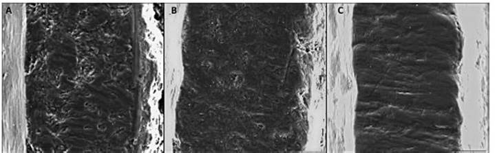
Fig. 1 Occlusal surface of different specimens of Lagostomus maximus at 400x magnification SEM. A, MACN-Ma 3928. B, MACN-Ma 3971. C, CFA-Ma 8706. Aand B have altered texture of the enamel surfaces and the original microwear features are indistinct or hard to distinguish. In C the enamel surface is unaltered and shows many scratch marks, a pattern usually seen on grazer mammals. Scale bar = 100μm.
For the purpose of this study, a tooth was selected for analysis from a skull of an adult Hydrochoerus hydrochaeris (Rodentia, Caviidae) (didactic collection, catalog number not as signed) from the mammal collection of MACN. This species was selected for the experiment as it was abundant in the didactic collection and because the chosen specimen presented no chemical treatment. These caviomorph rodents have continuously growing teeth (elodont), with enamel ridges parallel to dentin, meaning no oc clusal wear on enamel could expose dentin and lead to a microwear analysis in a structure other than enamel. Athird left upper molar was sepa rated and divided in three parts, where distal and mesial regions were treated with chemical com pounds (H2O2 and NaClO) and a third portion (central) remained with no treatments (control). The concentrations of hydrogen peroxide and sodium hypoclorite selected followed protocols indicated in the literature dealing with mammal collections (Díaz et al., 1998).
Molds of the occlusal surface of tooth samples were made before the treatments and after each interval of submersion using polyvinyl siloxane (President MicroSystem Light Body Coltène®), to track possible modifications on teeth microwear patterns. To obtain casts, the molds were then filled with liquid epoxy resin (CRISTALTACK ®) prepared with 2 parts of resin (component A) to 1 part of catalyst (component B) and let dry for 24 hours. Casts of teeth were mounted on pin stubs using cyanoacrylate adhesive and coated with 200 Å gold-palladium film using a sputter-coater ‘Termo VG Scientific SC 7620’. To evaluate the effects of each chemical compound, micropho tographies were obtained from the same region of the occlusal surface on control and treated samples using a SEMPhilips series XL model 30 at the MACNunder 20kV, 5mm distance work. There is not a standard magnification for mi crowear studies in rodents; so we defined a 400x magnification. This magnification was based on the width of the enamel ridges of Lagostomus maximus specimens; the same magnification was then applied to Hydrochoerus hydrochaeris.
Experiment 1
The distal portion of the tooth was treated following protocol indicated by Díaz et al. (1998): 1 minute of submersion in 1.25% solution of NaClO of domestic use (2.5%) and then sub merged in a 10% solution of H2O2 for intervals of 30, 60, 90 and 120 minutes. Díaz et al. (1998) were not clear about the interval of submersion in H2O2, so we tested different intervals to iden tify the effects of short and long exposures.
Experiment 2
The mesial portion of the tooth was sub merged in 1.25% solution of NaClO (2.5%) dur ing intervals of 30, 60, 90 and 120 minutes. The exposition intervals were selected by us knowing that intervals indicated in protocols are not cor rectly respected in many preparations and also considering the not neutralized reactions that al lows chemicals to continue acting even after the exposure intervals.
RESULTS
In Lagostomus maximus SEMimages (Fig. 1) it is possible to observe that specimens MACN-Ma 3928 and MACN-Ma 3971 have microwear features (scratches, pits and gouges) hard to distinguish by having an altered texture of the enamel surfaces. CFA-Ma 8706 presents an unal tered enamel surface and shows several scratch marks.
Experiment 1
In the sample treated with 1-minute NaClO and then with H2O2, the superficial scars of the enamel are progressively hard to distinguish, as their limits become more diffuse and there is a homogenization of the surface (see Fig. 2). For deeper structures it is possible to identify they become more visible under longer exposure time (e.g., gouge indicated by an arrow at left supe rior margin of the images at Fig. 2). At exposure intervals of 90 (Fig. 2 E) and 120 minutes (Fig. 2 F), the generation of a new pattern like paral lel scratches can be observed on occlusal enamel surface.

Fig. 2 SEMimages (100x100μm) of a third upper molar of Hydrochoerus hydrochaeris showing the enamel surface before treatments (A) and the changes in microwear patterns after a 1-minute submersion in NaClO (B) and different exposition intervals to H2O2: C) 30 minutes, D) 60 minutes, E) 90 minutes, F) 120 minutes. White arrows indicate the same surface structures changing along intervals. 400x magnification.
Experiment 2
The sample treated only with NaClO shows progressive changes in the size and shape of the scars (Fig. 3), resulting in a shift on the general morphology of the scars. At shorter intervals of submersion in this solution (30 and 60 min utes), there is a deepening of the scratches on the enamel and the entire teeth surface seems to be splitting into layers (Fig. 3 B and C). At higher exposure intervals (Fig. 3 D and E), the microwear patterns start to be obscured and there is an increased corroded surface, but it is possible to identify some new depressions with a gouge or pit morphology.

Fig. 3 SEMimages (100x100μm) of a third upper molar of Hydrochoerus hydrochaeris showing the enamel surface before treatments (A) and the changes in microwear patterns in different exposition intervals of NaClO: B) 30 minutes, C) 60 minutes, D) 90 minutes, E) 120 minutes. White arrows indicate the same surface structures changing along intervals. 400x magnification.
DISCUSSION
The preparation techniques of samples to be deposited in osteological collections has evolved along the years with the practice of cleaning with chemicals to remove tissues (e.g. sodium hypochlorite) and bleaching specimens using sodium hypochlorite and/or hydrogen peroxide (Díaz et al., 1998; Holden, 1914; Williams et al., 1977). Nowadays, maybe as a result of increased interest in natural history conservation (Cato et al., 2001), some authors recommend avoiding their use, applying it only if the specimen is for exhibition purposes and, even in these cases, just if it is really necessary (Hendry, 1999; Simmons & Muñoz-Saba, 2005).
Alterations caused by bleaching agents on dental material go beyond the superficial enam el, preventing teeth material to be used for many studies. The H2O2 is an oxidant agent with high capacity of forming free radicals like O2 and HO2 which can damage biomolecules preventing teeth to be used for DNAand other molecular stud ies (Hu et al., 1995; Ribeiro et al., 2006). Some studies concluded also that hydrogen peroxide and sodium hypochlorite alter isotopic values in bioapatite carbonate fractions, causing its disso lution (H2O2) and incorporating exogenous car bonates to it (NaClO) (e.g., Pellegrini & Snoeck, 2016).
There are different conclusions in dentistry research about the use of hydrogen peroxide concerning the effect on teeth enamel surface (Alqahtani, 2014; Joiner, 2007). Some works show it can alter the microhardness and super ficial texture of teeth enamel, due to demineral ization of the teeth (Jiang et al., 2008; Rodrigues et al., 2017). The effect seems also to be directly related to the concentrations and exposure time of the teeth to this bleaching agent (Bistey et al., 2007). This conclusion is supported by our re sults, where we identify that this chemical com pound not only deepens dietary scar present at the teeth but also generates a new micro relief at longer exposure time.
As in most biological collections the hydrogen peroxide used is generally the commercial one, the treated materials could be suffering the ef fects of not only hydrogen peroxide but also of the acids used to stabilize these solutions as more changes seems to be produced on enamel surface in an acid environment (Rodrigues et al., 2017; Xu et al. 2011). Commercial hydrogen peroxide generally has the addition of an acid to lower the pH values and ensure the stability of the product. The one we used, for example, has phosphoric acid, used on dentistry for acid etch ing and could be directly affecting the teeth sur face (Cerci et al., 2012; Kodaka et al., 1993). At longer intervals of exposure to H2O2, 90 and 120 minutes, some parallel grooves become evident, which could reflect the alignment of the enamel microstructure that becomes visible after the re moval of the superficial enamel (Koenigswald & Clemens, 1992).
Sodium hypochlorite is also used in dentist ry but not as a bleaching agent, as in the case of hydrogen peroxide, but for teeth steriliza tion and deproteinization (Amaechi et al., 1998; Ekambaram et al., 2017). When applied on teeth, NaClO increases dentinal tubules and enamel pores, turning the tissue more permeable and enhancing, for example, the bond between teeth and resins (Abdelmegid, 2018; Silva et al., 2018). It is possible to see in our images, mainly at lon ger exposure times, that NaClO deepens many scars and creates new ones mainly with a gouge or pit aspect, what could be a result of enhanced corrosion due to larger permeability and a larger surface exposed to the action of this chemical compound.
The diversity of textures observed on altered samples is a combination of the chemical com pound and the exposure intervals chosen. For shorter intervals (30-60 minutes), H2O2 seems to cause fewer modifications on general teeth microwear pattern when compared do NaClO. Any case, it is crucial to notice that even shorter periods of submersion in any of these chemical compounds altered the original microwear pat tern of the teeth, disabling irreversibly the treat ed materials for microwear studies as they affect natural wear patterns produced by specimen dietary behaviors, leading to erroneous dietary inferences when using these samples.
CONCLUSIONS
The chemical attack by H2O2 and NaClO used for cleaning and bleaching skeletons and skulls continues to be used in some biological collec tions, even when its negative impacts were pre viously adverted for DNAand isotopic studies. It is important to highlight here that the chemical compounds, mainly sodium hypochlorite, may not be neutralized when the skulls and skeletons are washed with water as this procedure only di lutes the compounds, meaning the chemical can continue acting a longer period even on a lower concentration. Specimens in biological collec tions are stored by many years, so time plays an important role on materials treated with bleach ing agents and the effects of this longer exposure has to be studied as a particular case when com pared to dentistry research.
Summing up, we show the effects of H2O2 and NaClO on teeth surface with special attention to dental microwear, bringing evidence for the use of alternative preparation and conservation tech niques that allow the recovery of information. New experiments in other groups and using dif ferent concentration and/or exposure times for any chemical compound used in biological col lections could contribute to the knowledge about the effects of some practices on osteological and teeth samples.

