


 Inglés (pdf)
 Articulo en XML
 Referencias del artículo
 Enviar articulo por email
 Citado por SciELO
  Similares en
SciELO
Similares en
SciELO 
 Permalink
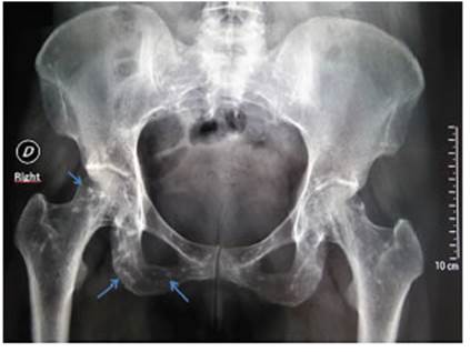
A 40-year-old female patient, presented to the Emer gency Department after suffering minor trauma. She de nied any significant past medical history. Different x-rays showed multiple well circumscribed, ovoid, radiodense bony lesions in the shoulders and pelvis, consistent with the diagnosis of osteopoikilosis (arrows in Figures 1 and 2). Subsequently, she provided previous x-rays, where the described images could be observed.
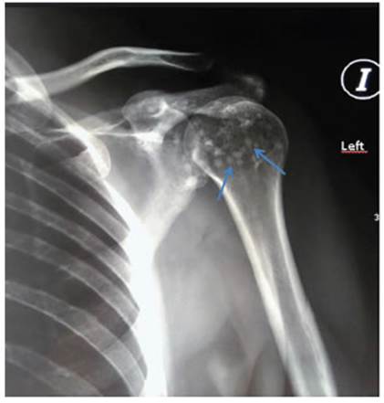
Osteopoikilosis is a rare, benign, autosomal dominant disorder characterized by sclerotic bone lesions most commonly involving feet, hands, pelvis, and metaphy sis of long bones. Appearances on imaging are those of multiple, small well-demarcated 2-3 mm circular or ovoid radiodense, sclerotic lesions. Histologically, the sclerotic areas are focal condensations of compact lamellar bone within the spongiosa. Osteopoikilosis is caused by a mu tation in the LEMD3 gene and is usually clinically asymp tomatic and typically detected on incidental imaging.
The symmetric distribution, lack of bone destruction, and location differentiates osteopoikilosis from meta static disease, which tends to be seen more often in ribs, vertebral bodies, and the diaphysis of long bones.

