


 Inglés (pdf)
 Articulo en XML
 Referencias del artículo
 Enviar articulo por email
 Citado por SciELO
  Similares en
SciELO
Similares en
SciELO  uBio
uBio 
 Permalink
TEXTO COMPLETO
Bacillus thuringiensisis a Gram-positive and sporulated bacterium exhibiting insecticidal activity against a wide range of insects.3During sporulation, this bacterium produces a number of different proteins forming crystalline inclusions adjacent to the spores (parasporal crystals). Among these insecticidal proteins, the most abundant are those commonly known as Cry (Crystal) proteins, which are responsible for exerting a toxic activity (upon ingestion) against insects of different species.5For this reason,B. thuringiensishas proved to be the most efficient and used bioinsecticide to date.2However,Spodoptera cosmioides,Spodoptera eridaniaandAgrotissp. (Lepidoptera) are species that are not yet controlled by some transgenic crops (e.g. Intacta RR2Pro soybean). Thus, in an attempt to enlarge the host spectrum of this bacterium it is necessary to search for novel strains. In this work we show a sporulatedB. thuringiensisBt-UNVM_84 strain exhibiting a number of rare amorphous to spherical crystal combinations, whereas sporulatedB. thuringiensisstrain Bt-UNVM-94 showed quasi symmetric bipyramidal parasporal crystals, by using scanning electron microscopy (SEM) (Fig. 1). Strains Bt-UNVM_84 and Bt-UNVM_94 were isolated from Oncativo (Córdoba, Argentina) and Cululú (Santa Fe, Argentina), respectively. The insecticidal activity of these differentB. thuringiensisstrains is currently under investigation. Each strain was grown in liquid CCY sporulation medium6for ∼48h (150rpm) until no vegetative cells were observed under a light microscope. The presence of parasporal crystals was first determined using Coomassie blue stained slides1(1000×) under a Nikon E100 light microscope and confirmed later by a NikonTi-Eclipse phase contrast microscope (1000×) (data not shown). For the SEM analysis, aliquots of 1ml were centrifuged for 5minutes (16,000g) at room temperature. Each pellet was washed three times with sterile distilled water and fixed with 100μl 4% formaldehyde. Each fixed preparation was then sent to Centro Integral de Microscopía Electrónica (CIME - CONICET - UNT) for SEM examination.
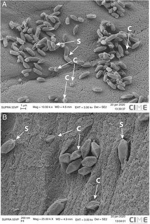
Figure 1: Scanning electron microscopy of parasporal crystals fromBacillus thuringiensisstrains Bt-UNVM_84 and Bt-UNVM_94 (C=crystal, S=spore). (A)B. thuringiensisstrain Bt-UNVM_84 showed combinations of amorphous to spherical parasporal crystals of ∼0.7-0.9μm from two points along the diametral axis. (B)B. thuringiensisstrain Bt-UNVM_94 exhibited quasi symmetric bipyramidal parasporal crystals of ∼1.0-1.2μm from two points along the longitudinal axis. Crystal size was measured using ImageJ.4Parameters used for image acquisition are shown: Mag=magnification (K×=1000×), WD=work distance, Eht=energy high tension, Det=detector type and SE2=secondary electron. (0,52MB).
Funding
This work was supported by Universidad Nacional de Villa María research grant PIC UNVM 2018-2019.
ACKNOWLEDGMENTS
We thank to Hernán Esquivel, Luciano Martínez from CIME-CONICET-UNT and Vanessa Areco for their technical assistance. We also thank to the Instituto de Investigación of Universidad Nacional de Villa María for contributing to this ongoing research.

