


 Español (pdf)
 Articulo en XML
 Referencias del artículo
 Enviar articulo por email
 Citado por SciELO
  Similares en
SciELO
Similares en
SciELO 
 Permalink
Introduction
Edematous conditions affecting pregnancy are uncommon during late gestation in domestic species (Momont 2005), being more frequent in cattle (Schlafer and Foster 2016). The excessive accumulation of fluid in either the amniotic or the allantoid cavities, termed respectively hydramnios and hydrallantois, are the most frequent causes of dropsy in cattle (Youngquist and Threlfall 2007), reaching over 90% of the dropsy cases. In general, both occur separately as distinct entities, but occasionally, the involvement of both compartments can be seen (Schlafer and Foster 2016). Other dropsical conditions include edema of the chorioallantoic membrane or edema within the fetus, such as fetal anasarca, fetal ascites and hydrothorax (Roberts 1986).
Normally, the origin, quantity and nature of the fluid cavity change throughout the normal gestation (Schlafer and Foster 2016). Amniotic fluid is clear and slightly viscous, and it is mostly derived from the salivary secretion of the fetus, then, its volume is largely depending on fetal swallowing. For this reason, the occurrence of hydramnios is always associated with fetal abnormalities, usually malformations (Drost 2007, Schlafer and Foster 2016). In contrast, allantoic fluid has a clear, watery consistency, and it is largely produced by excretion from the fetal kidneys (Momont 2005). Excessive accumulation of allantoic fluid is attributed to placental pathology, and it is much more frequent than hydramnios (Wintour et al. 1986, Drost 2007). Among the various hydropic conditions that can affect a pregnant cow, this is the sole condition that qualifies as a genuine emergency with favorable response when treated (Momont 2005).
Hydrallantois is associated with a decrease in the number of caruncles, and although the origin of this reduction remains undetermined (Youngquist and Threlfall 2007), transplacental poisoning and fetal damage leading to hydrallantois associated with the ingestion of Sida Carpinifolia in cattle has been described (Reis et al. 2019). It is frequently associated with non-functional caruncles in one of the uterine horns, and enlargement of the remaining placentomes (Drost 2007, Schlafer and Foster 2016). The remaining caruncles tend to show hypertrophy. The fetal membranes are usually only slightly thickened, although this is not always the case (Schlafer and Foster 2016).
The aim of this paper is to describe two cases of hydrallantois in beef cattle from the province of Buenos Aires, Argentina, including the associated gross and microscopic findings. This work also aims to appoint the importance of reaching a diagnosis in cases of acute and subacute death in cattle.
Materials and Methods
The first case (Case 1) took place on a beef cattle farm located near Azul, province of Buenos Aires. The herd consisted of 250 Aberdeen Angus, cows that came from Santa Fe province five months earlier (May, 2018). The animals were grazing on natural pasture. The sanitary plan included vaccination against Clostridium chauvoei, Cl. septicum, Cl. perfringens types C and D, and Escherichia coli, parenteral administration of copper and oral supplementation with mineral salt blocks, including magnesium. Calving had started during the month of July, and upon consultation four animals had been found dead during the past month. In July a necropsy was performed on an adult Aberdeen Angus cow with approximately 12- 15 hours postmortem. Autopsy was performed and samples of liver, heart, lungs, spleen, kidneys, uterus, and placenta were collected for histopathological analysis.
The second case (Case 2) took place on a beef cattle farm located in Saladillo, province of Buenos Aires. The herd consisted of 151 red Aberdeen Angus cows. The animals were grazing on natural pasture and the sanitary plan included a double pre-breeding dose of vaccine against reproductive diseases (Bovine herpesvirus type 1, Bovine viral diarrhea virus, Campylobacter fetus, Histophilus somni and Leptospira interrogans). The cows had been inseminated in October 2017, and calving had started during mid-June 2018. The problem started with a cow exhibiting bilateral abdominal distention beginning at the eight months of gestation. An excessive amount of fluid was noted on rectal examination, preventing palpation of fetal structures. Fifteen days later, the animal was found lying in ventral recumbency and unable to stand. The cow died three days afterwards, and a necropsy was performed. Necropsy was performed and samples from placenta, including placentomes and interplacentomal area, were collected for histopathological analysis.
The samples for histopathological analysis from both Case 1 and Case 2 were fixed in 10% neutral buffered formalin (pH 7.2) for 48h. Samples were then dehydrated with ethanol and embedded in paraffin to obtain blocks which were then sectioned by means of a rotary microtome to 6-μm-thickness sections. They were then rehydrated in distilled water and stained with hematoxylin and eosin (H&E).
Results
The animal from case 1 had been found dead, lying in sternal recumbency with its hindlimbs extended caudally, interpreted as an acute death (Figure 1).
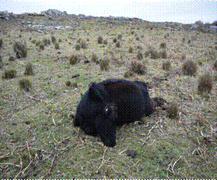
Figure 1 Case 1, Aberdeen Angus, pregnant cow. The animal was found lying in sternal recumbency with its hindlimbs extended, indicating a sudden death.
On external examination, the abdomen was markedly bilaterally distended. At necropsy, when the abdomen of the cow was opened, the abdominal musculature was separated by large quantities of a yellow, viscous material compatible with edema and both uterine horns were observed to be distended to more than 15 times their normal size. The incision of the right (non-pregnant) horn resulted in the exit of about 50 to 70 liters of allantoic fluid (Figure 2). The left horn also exhibited a large volume of allantoic fluid. The uterus was gravid with a full-term fetus and did not show any gross abnormalities.
The cotyledons had a thickness of 2 to 3 cm. The placentomes were pedunculated and measured 7 cm in diameter, and 2.5 to 3 cm deep. No other macroscopic lesions were observed.
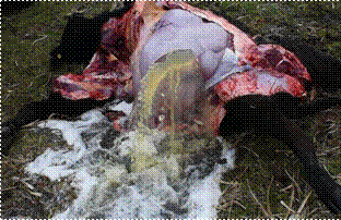
Figure 2 Case 1, Aberdeen Angus, pregnant cow. Note the abundant amount of allantoic fluid coming out from the right uterine horn.
In Case 2, grossly the left uterine (pregnant) horn was distended to more than 10 times its normal size (Figure 3). After incision, about 80 liters of clear watery fluid came out of the allantoic sac. The uterus was gravid with an 8-month fetus. A total of 55 placentomes were counted, 7 of which were inspected. These placentomes were pedunculated and measured 8 to 10 cm long, 3 to 4 cm wide, 4 to 5 cm deep and a weight between 133 and 140 g. When incised below the chorioallantoic arcade in the region of crypts/villi, some placentomes exhibited multiple coalescing hemorrhagic foci. The interplacentomal area showed severe, diffuse oedema. The maternal chorion was firmly adhered to the fetal part of the placentome, and separation using manual traction was difficult. When detached was achieved, numerous crypts were noted to be dilated, and some of them had remnants of chorionic villi. In the umbilical cord, both the vessels and the allantoic duct were distended. No other macroscopic findings were present in either the cow or fetus.

Figure 3 Case 2, Aberdeen Angus, pregnant cow. Marked distension of left uterine horn with abundant allantoic fluid.
In both cases, the microscopic lesions observed in the placenta were similar and included separation of the fetal trophectoderm by evident regions of connective tissue within the placentomes, and edematous caruncular stroma (Figure 4 A). Extensive foci of mineralization, occasionally associated with the absence of chorionic villi and uterine epithelium, disorganized villi with low or absence of vascularization (immature chorionic villi), and mild diffuse congestion of both the maternal and fetal endothelium were also present (Figure 4 B). Only in Case 1 samples other than placenta were collected, observing marked diffuse cytoplasmic vacuolation in hepatocytes, indicative of hydropic degeneration, along with the loss of hepatocyte cords. The remaining organs, i.e., the heart, lungs, kidneys and spleen, did not exhibit any relevant microscopic lesions.

Discussion
Hydrallantois has been described in both dairy and beef cows. Typically, allantoic fluid volume increases steadily from late gestation to roughly 10 L, ranging between 8 and 15 L (Drost 2007). In cases of hydrallantois, this increase is usually rapid, progressing over 5 to 20 days (Pickles 1987). The fluid composition closely resembles plasma, with markedly higher sodium, potassium, chloride and creatinine levels than normal allantoic fluid (Youngquist and Threlfall 2007). According to the situation Momont (2005) described for hydrallantois, the cases reported in this work exhibited large amounts of allantoic fluid, nearing roughly 150 L. Although only acute death was observed in Case 1, Case 2 exhibited a subacute course allowing for clinical signs to be observed. Sudden death of Case 1, we believe occurred by compression of thoracic cavity causing asphyxiation, and impediment of normal systemic circulation, as occurs in bloat (Yirdachew and Mekonnen 2022).
The presence of multiple fetuses, in vitro fertilization, and transgenic and cloning technologies have been associated with an increased risk of developing this condition (Youngquist and Threlfall 2007, Schlafer and Foster 2016), although these findings were not observed in the cases described here. Bovine viral diarrhea virus has also been implicated in cattle hydrallantois cases (Collyer 1990). Placental abnormalities found in the presence of hydrallantois, such as reduced vascularization, particularly at the apex of the villi and loss of differentiation of the trophoblastic epithelium (primitive villi), suggesting that an insufficient development of villi could contribute to the physiopathology of this condition (Loi et al. 2006, Feliciano et al. 2013).
Hydrallantois is associated to diseased uterus in which most caruncles in one horn are not functional with consequent reduction of placental vascularization resulting in metabolic changes thereby accumulating fetal fluid (Drost 2007, Kapadiya et al. 2018). The 2 cases presented herein had low number of placentomes with increased size, low vascularization and inadequate development (immature villi), which is a compensatory mechanism during gestation, of congenital origin with inadequate uterine development, as no inflammatory findings were present. Consequently, edema formation due to low vascularization leads to further hypoxia causing an increase in vessel permeability and interstitial fluid accumulation, leading to insufficient transplacental exchange rate and accumulation of fluid in the allantoic cavity (Dini et al. 2020). It has also been associated with a recessive fetal abnormality manifested with fetal polyuria and subsequent hydrallantois (Sasaki et al. 2016). Even though no genetic and hereditary factors were evaluated in the cases described here, the fetus from both cases did not exhibit gross renal abnormalities. Findings such as at-term fetuses exhibiting ascites and an enlarged umbilical cord are frequent findings associated to placental disfunction, including hydrallantois (Constant et al. 2006, Schlafer and Foster 2016). Although no fetal abnormalities were observed grossly in Case 1, the fetus from Case 2 did exhibit both ascites and an enlargement of the umbilical cord. Although the cause of the edema within the placentomes is unknown, we believe it could be directly associated with hydrallantois, as fetal membranes play an important role in regulating the composition and volume of fetal fluids (Wintour et al. 1986).
Gross and microscopic findings in cases of hydrallantois include an inadequate number of caruncles and adventitial placentation. The remaining caruncles tend to be hyperthophied, and fetal membranes are usually slightly thickened (Schlafer and Foster 2016). In both cases, placentomes were slightly enlarged compared to previous reports on normal placentae. Normal size during late gestation is 5 to 8 cm diameter (Laven and Peters 2001, Barbeito 2010, Kouamo et al. 2018), 2 to 3 cm width (Schlafer et al. 2000) and 3 cm depth (Ribeiro et al. 2008), and an individual weight between 51 and 114 g (Laven and Peters 2001, Constant et al. 2006, Adeyinka 2012). Some works report placentomes with a diameter of 12 cm (MacDonald 2011). Despite the severity of the inflammatory lesions and mineralization in Case 2, these findings have been described in normal placentae and can thus be considered incidental (Schlafer and Foster 2016). Hydropic degeneration of hepatocytes found in Case 1 may be produced by anorexia due to prolonged discomfort or recumbency.
The diagnosis of hydrallantois in cattle can be made by rectal palpation of a distorted uterine horn without palpation and ballottement of the fetus and the placentomes (Bhattacharyya et al. 2012). However, as seen in Case 1, the course may be too acute to enable the diagnosis employing rectal palpation. A rapid bilateral abdominal distention history in the last 15 to 20 days suggests hydrallantois. If a C-section is performed, confirmation is made by observing large quantities of clear allantoic fluid (Bhattacharyya et al. 2012). In both of the cases presented here, a diagnosis of hydrallantois was made post-mortem based on the origin of the fluid within the uterus, the presence of at-term fetuses lacking gross and microscopic abnormalities (although one of them exhibited ascites), and the presence of edema within the intercotyledonary region and the placentomes. We hypothesized that the underlying factor leading to the demise of Case 1 was acute cardiorespiratory failure resulting from heightened intraabdominal pressure from the enlarged uterus. It is unlikely that this condition was the cause of death of the animals that had died previously in the herd, but no necropsies were performed in those cases. In Case 2, only one animal developed hydrallantois, and though treatment is favorable for these cases, diagnosis was not proper when still alive, not reaching for treatment. The most common treatment for hydrallantois is termination of pregnancy by means of cloprostenol or other prostaglandin F2α analogs. Induction with corticosteroids and estrogen preparations has also been reported (Sharp et al. 1978). However, delivery by C-section is the preferred choice when the animal may not withstand the stress of natural parturition (Bhattacharyya et al. 2012). Particularly, early hydrallantois detection is essential as applying treatment with favourable results.
Conclusion
In Argentina, several acute deaths among adult cattle occur without reaching a definitive diagnosis or simply because of a lack of diagnosis, with no official data available and numerous cases remaining undiagnosed. Because of this, we encourage performing a full diagnosis work-out in all acute or sub-acute death cases to find other common or uncommon conditions that could affect herds of beef cows. Though unusual, hydrallantois should be considered a differential diagnosis for acute death in beef cows, including hypomagnesaemia, anthrax, bacillary hemoglobinuria, nitrate intoxication and plant poisoning.

