


 Inglés (pdf)
 Articulo en XML
 Referencias del artículo
 Enviar articulo por email
 Citado por SciELO
  Similares en
SciELO
Similares en
SciELO 
 Permalink
A little history
Asthma and Chronic Obstructive Pulmonary Disease (COPD) are heterogeneous, obstructive airway diseases, whose physiopathology is far from being completely understood even today.
Over 60 years have passed since the almost simultane ous generation of two hypotheses regarding the genesis of asthma and chronic bronchitis associated with COPD. Despite the time that has elapsed, with sometimes pas sionate and personal conflicts, they have generally been presented as opposing hypotheses: the assertion of one denied the other, and vice versa.1-8
More than six decades have passed since then, and the objective of this manuscript is to review which concepts have been confirmed over time in the light of recent research which, in the opinion of the author of this manuscript, have been relevant.
What did the British hypothesis say?
In the early 1950s, COPD as such was not yet described, and Lynne McA Reid and McLean as sociated smoking with the presence of bronchor rhea, chronic cough, changes in bronchial mucosal defense, bacterial colonization, and frequent infec tions that led to the genesis of chronic obstructive bronchitis. This hypothesis was known as the “British hypothesis” (Figure 1).1,2
What did the Dutch hypothesis say?
Since asthma and COPD share some common aspects, between 1961 and 1964, Dr. NG Dick Orie proposed in his doctoral thesis at the University of Groningen, Neth erlands, that asthma, chronic bronchitis, and emphysema were phenotypic expressions of the same disease and that they evolved from one to another as individuals aged, influenced by different factors: “Bronchitis and asthma may be found in one patient at the same age but as a rule there is a fluent development from bronchitis in youth to a more asthmatic picture in adults, which in turn develops into bronchitis of elderly patients” (Figure 2).3,4

As a result of the interest generated by such a bold hypothe sis, an International Symposium on “Chronic Bronchitis” was organized in 1962.5 The relationship between exogenous factors (environment, exposure to allergens, and tobacco smoke) and endogenous factors (atopy and bronchial hyperreactivity) would express itself differently in chronic bronchitis. This hypothesis has caused a significant debate among researchers from the United Kingdom, the rest of Europe, and the United States of North America until recent years.5-10 In 1969, Dr. Fletcher labeled it as the “Dutch Hypothesis”.
MORE THAN 60 YEARS LATER, WHAT NEW INFORMATION IS AVAILABLE ABOUT THE BRITISH HYPOTHESIS?
When the British hypothesis was formulated, sev eral facts were unknown, or weren’t sufficiently known as they are today. These include, for ex ample, the evolution of the concept of pre-COPD, the importance of the presence of respiratory symptoms in early stages of COPD, the charac terization of the frequent exacerbator phenotype, and the importance of respiratory microbiota. All of these factors currently strengthen what was stated more than 60 years ago by Lynne McA Reid.1
Chronic respiratory symptoms
While the fundamental work of Fletcher et al showed that chronic bronchitis (-CB- chronic cough and chronic bronchorrhea) and chronic airflow obstruction were two separate clinical conditions that could be associated and were not related to an accelerated loss of the lung function, more recent studies have revisited this concept, leading to the proposal of the “Pre-COPD” stage, which will be discussed in the following section. Bronchorrhea is associated with a greater decline in the forced expiratory volume in the first second (FEV1) and a higher risk of developing COPD in young smok ers with CB.11-14 It is also associated with a higher number and greater severity of exacerbations.15 Several cohort studies have examined the pres ence of chronic respiratory symptoms and their relationship with the progression towards COPD in individuals with preserved lung function.16-22 In the SALPADIA-1 study, it was found that indi viduals with bronchorrhea and nearly normal lung function (GOLD 1 [Global Initiative for Chronic Obstructive Lung Disease]) experienced a higher decline in FEV1 and increased use of healthcare resources over a 3-year follow-up period.16 In the Copenhagen City Heart Study, it was determined that over a 5-year period, the odds ratio (OR) for the presence of bronchorrhea as a risk factor for COPD was 1.1 (0.9-1.4), and over a 15-year period, it was 1.2 (0.9-1.6). However, bronchorrhea was associated with a greater decline in FEV1 and increased morbidity (hospitalization, OR of 5.3 for men [2.9-9.6] and 5.1 for women [2.5-10.3]).17 In the SPIROMICS cohort, it was determined that smokers with normal lung function already exhibited increased inflammatory cellularity in bronchial mucosa compared to controls.18 In the COPDGene cohort, an accelerated decline in lung function was observed in smokers with normal lung function.19 In the UK Biobank cohort, 351,874 subjects were studied for 9 years, examining the relationship between airflow obstruction and the presence of respiratory symptoms.20 Among other factors, it was determined that the deteriora tion in the lung function was strongly associated with the presence of respiratory symptoms and cardiovascular comorbidities (adjusted OR of 2, 95% confidence interval [CI] 1.91-2.14, p<0.0001, and 1.71 [1.64-1.83], p<0.0001).20 Lung function deterioration was associated with overall mortal ity (hazard ratio [HR]1.61 [95% CI 1.53-1.69], p <0.0001) compared to controls.20 In a study of Sherman et al of 3,948 subjects studied for 12 years, which compared patients with and with out respiratory symptoms (persistent wheezing, chronic cough, chronic expectoration, or dyspnea) in relation to FEV1 and adjusted by exposure to tobacco and height, it was observed that men with chronic cough and women with chronic expectora tion exhibited an accelerated FEV1 decline.21 In a Copenhagen study by Lange et al, involving 13,756 subjects studied for 10 years, it was determined that chronic expectoration was weakly associated with overall mortality (relative risk [RR] of 1.1 for women and 1.3 for men). However, in those with severe obstruction (FEV1 of 40%), the risk was much higher (RR of 4.2).22 Currently, the therapy recommended by the GOLD guidelines is based on the ABE matrix classification, considering the presence of dyspnea, the degree of impairment in the quality of life, and the type and number of exacerbations.23 However, at present, the presence of chronic bronchorrhea or chronic cough or the degree of bronchial obstruction are not considered in therapeutic decisions. FEV1 is an independent factor for mortality and has been used as an in clusion criterion for the clinical development of current long-acting bronchodilators and inhaled corticosteroids, as well as their combinations in the last 20 years.24 Anyway, recent research shows, as an example, that patients classified as GOLD A or B with mild or severe bronchial obstruction don’t have the same disease progression. But this is not taken into account by the current pharmacological treatment recommendations of the GOLD guide lines.25-26 A recent study by Han et al in individuals with tobacco exposure (>10 pack-years) and a CAT score >10 (respiratory symptoms) showed that dual long-acting bronchodilator therapy does not improve the quality of life.27
Pre-COPD concept
All of the historical information mentioned above has recently been taken up by a group of renowned international specialists who published a document where they propose to reconsider the controversial “Stage 0” concept from the 2001 GOLD guidelines and replace it with “Pre-COPD” for patients who do not meet the current GOLD criteria for COPD. This is based on three domains:28-30
A. Clinical symptoms: presence of bronchorrhea, cough, dyspnea and exacerbations.
B. Functional: patients with a post-bronchodila tor FEV1/FVC ratio greater than 0.7 but with signs of air trapping in lung volume measurements, reduced DLCO (carbon monoxide diffusion), or signs of small airway obstruction.
C. Imaging: presence in chest computed tomog raphy of centrilobular emphysema, thickening of the bronchial walls of the large airway, or signs of involvement of the small airway.
Microbiota and respiratory diseases
The community of microorganisms including bacteria, fungi, viruses, and archaea that inhabit our body collectively constitute the “human mi crobiota”. If we consider the entire genetic load of these microorganisms, it is referred to as the “human microbiome”.31-33 The airways aren’t ster ile, and a community of microorganisms resides there, interacting with our body in a balanced manner in a healthy state. The lower airways have a lower biomass of microorganisms due to fewer nutrients and local immuno mucociliary clearance mechanisms.31-33 Microorganisms can enter from the oropharynx through micro-aspirations or dis persion through the mucosa.31-33 As a result, the respiratory microbiota has a direct interaction with the gut microbiota, especially that of the upper airway.31-33 The interaction between both microbiotas also occurs systemically through various metabolites of the intestinal bacteria that affect the systemic immune system. This interaction involves not only bacteria (which are the most studied) but also the mycobiome and pulmonary virome.31-33 When there is an imbal ance in this host-microorganism interaction, it is referred to as “dysbiosis”.31-35 Dysbiosis can be caused by antibiotics, nutritional disruptions, or external infections that alter the benign resident commensal flora. There has been increasingly solid evidence every year for the past two decades that the alteration of the microbiome plays a role in several diseases.31-35 This applies to airway dis eases such as asthma, COPD, or cystic fibrosis, and to other conditions traditionally considered unrelated to microorganisms, such as idiopathic pulmonary fibrosis, cancer, or adult respiratory distress syndrome.31-35 In the case of asthma, exposure to microorganisms at an early age has long-term consequences in susceptibility.36
The first generation of studies focused on de scribing the genetic sequence of 16S rRNA to char acterize the microbiotas of the digestive and respi ratory tracts. Following in vitro and in vivo studies in animals, controlled studies in humans began to assess the host-microorganism interaction in diseases. Some studies of multicenter academic consortiums are currently being developed to ac count for population and geographic variability, which can influence the findings, as well as method standardization and multi-omics data analysis, going beyond bacteria and including viruses, fungi, and archaea. Additionally, it’s important to consider inter-individual variability in the course of the disease and the response to various treat ments. We are at the start of a new era in precision medicine where the microbiome could contribute to the understanding of new disease pathogenesis, diagnoses, and treatments.32 COPD is a complex syndrome characterized by different phenotypes, all sharing the common feature of chronic airflow obstruction. The microbiota in COPD substantially differs from that of healthy control individuals, and this difference is even more pronounced during exacerbations.35 The dynamics of these changes are influenced by multiple factors, including the phenotypic heterogeneity of COPD, physiopathological changes, treatments (such as corticosteroids and antibiotics), smoking, and exacerbations.35 Approx imately 40 to 50% of exacerbations are triggered by bacteria that increase airway inflammation and obstruction, as well as bronchorrhea. The most frequently involved bacterial genera are Strepto coccus, Pseudomonas, Moraxella, Haemophilus, Neisseria, Achromobacter, Corynebacterium, and atypical bacteria like Mycoplasma pneumoniae and Chlamydia pneumoniae.35 Particularly with regard to COPD, the bacterial load is related to a higher incidence of exacerbations and a decline in lung function.36 A specific strain can generate a specific immune response, and the appearance of a differ ent strain increases the risk of exacerbations.37 Bafadhel et al identified fundamental differences in immunotherapy.38 The type of exacerbation can be predicted during the stable phase of COPD. In cases of frequent exacerbations, the pattern tends to repeat itself. The bacterial phenotype was found in 55% of the cases, the eosinophilic in 29%, viral in 28%, and the remainder were pauci-inflammatory. IL-6 and IL-8 levels can predict among frequent exacerbators which are the more prone to exacerbate.38 Additionally, viral infec tions can disrupt microbiome balance, increasing susceptibility to secondary bacterial infections and associated exacerbations.31-35 Respiratory syncytial virus, influenza A virus, and rhinovirus infections increase the expression of bacterial adhesion molecules (e.g., ICAM-1, PAFR, CEACAM-1) on epithelial cells, promoting the development of H. influenzae, S. pneumoniae, and P. aeruginosa.39-40 Respiratory viruses also deteriorate mucociliary clearance and damage epithelial cells, disrupting the first line of defense in the respiratory tree mucosa and allowing the invasion of pathogenic bacteria through it.39-40 Interestingly, it was ob served in animal models that the relationships between the gastrointestinal tract microbiome and metabolites produced by commensal bacteria in the digestive tract protect against respiratory virus infections, while those produced by the re spiratory microbiome protect against bacterial and viral infections.39-40 Fungi such as the Aspergillus genus have been identified as etiological factors in the exacerbations of asthma, COPD, cystic fibrosis, and bronchiectasis.31
Respiratory infections and exacerbator phenotype
A history of severe childhood infections is as sociated with decreased lung function and the presence of respiratory symptoms in adulthood.41 There is evidence that infection with the human immunodeficiency virus (HIV) represents an in creased risk of developing COPD (OR 1.14, 95% CI 1.05-1.25), as well as tuberculosis.42-43 Starting with the work of Soler Cataluña et al which clearly demonstrated that having 1-2 exacerbations in the previous year, or even more, compared to not having any, presented an increased risk of mortality and hospitalization.44 Donaldson et al showed that the subgroup of patients who experi ence frequent exacerbations also experience an accelerated decline in their lung function.45 Furthermore, all of this was associated with a poorer quality of life.46 The ECLIPSE study provided additional information about how the subgroup of patients who experienced frequent exacerbations in the previous year were more likely to continue exacerbating over three years of follow-up, while the opposite occurred in those who had never experienced exacerbations before.47 Since 2011, the GOLD guidelines have considered this condition as a phenotype that allowed rating the higher risk of morbidity and mortality and conditioning the specialized pharmacological treatment.23
MORE THAN 60 YEARS LATER, WHAT NEW INFORMATION IS AVAILABLE ABOUT THE DUTCH HYPOTHESIS?
Many advances in the field of genetics in asthma and COPD, the impact of neonatal development in lung function, exposure to biomass smoke, the presence of bronchial hyperreactivity in COPD, the eosinophilic exacerbator phenotype in COPD (or asthma-COPD overlap), and the eosinophil as a biomarker, have or would have implications in current management. These advancements have strengthened what was originally proposed as the “Dutch hypothesis” more than 60 years ago by Dick Orle.3-10
Advances in genetics in asthma and COPD
The revolution in genetic research has been one of the most marvelous and rapid advancements in understanding the physiopathology and etiology of many diseases, including asthma and COPD, in the last twenty years since the complete development of the Human Genome Project.
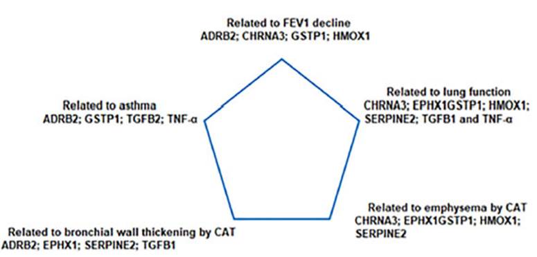
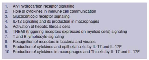
In 2011, Dirkje Postma revisited the Dutch hypothesis in view of the advances in genetics and environmental factors common to both asthma and COPD.48 Based on genetic load, various en vironmental factors (allergens, irritants, tobacco, etc.) triggered different rates of fetal lung tissue growth. After birth, the relationship between genetics and environmental factors (epigenetics) allowed for the expression of different clinical phenotypes (Figure 3).48 More than ten years after the formulation of the “Dutch hypothesis”, Fletcher and Peto identified in their famous study a group of individuals who, despite exposure to tobacco smoke, would not develop COPD (they called them “non-susceptible”), while others would (“susceptible”).11 Kaneko et al reviewed the list of coding genes common to the development of asthma and COPD.49 At least ten molecular signaling pathways have been determined in the associated genesis of asthma and COPD, each related to several regulator genes (Table 1).49 More recently, Agusti and Hogg summarized the 22 genes that are most closely associated with the development of COPD.50 These are: TGFB2, PID1, RARB, EEFSEC, FAM13A, GSTCD, HHIP, TET2, DSP, HTR4, ADAM19, AGER, ADGRG6, ARMC2, SFTPD, RIN3, THSD4, CHRNA5, CCDC101, CF PDP1, MTCL1 and CYP2A6. Some of these genes are related to another famous theory that also explains part of the physiopathology of COPD: the “proteases and antiproteases” theory.23,39,40,50 Since the last century, the action of proteases and the destruction of pulmonary elastic tissue have been related to emphysema.23,39,40,50 The main proteases are neutrophil elastase and proteinase-3. Serine proteases are potent stimulators of mucus production and induce bronchorrhea in patients with chronic bronchitis. More recently, it has been determined that matrix metalloproteinases (MMPs) MMP-1 and MMP-9 derived from macro phages and neutrophils, they are overexpressed in patients with emphysema and their synthesis is induced by the action of tobacco.23,39,40,50 However, normal lung tissue is protected against them by the activity of the antiproteases. The most significant inhibitor of serine proteases is the alpha-1 anti trypsin protein, an alpha-1 globulin. The genetic model of emphysema caused by alpha-1 antitrypsin deficiency has been extensively studied, and cur rently it is possible to diagnose it early and treat it with replacement therapy using the protein.23,39,40,50 Another genetically significant factor is the shortened length of telomeres, which is related to increased susceptibility of the emphysema.51-52 Morla et al demonstrated in an interesting con trolled study involving normal individuals that telomere length shortens in smokers (p = 0.05), especially those with a higher smoking load (p < 0.001), and in the presence of bronchial ob struction.52 It has even been determined which are the mutations in the telomerase-regulating gene that have a higher risk of emphysema, idio pathic pulmonary fibrosis, primary bone marrow failure, and hair loss, and these are inherited with autosomal dominance.52
Neonatal development and lung function
The trajectory of lung function growth is es tablished from gestation, birth, childhood, and adolescence.48,53-54 50% of patients who develop COPD may not be associated with accelerated loss of the lung function but rather to abnormal lung growth during gestation and early childhood.55 Genes involved in lung development, together with maternal exposure to tobacco or biomass smoke, have significant influence on the develop ment of asthma and COPD.48,56-57 The expression of different genes during the development of the uterine airway, such as Wnt gene signaling, has been associated with decreased lung function in childhood and asthma.58-59 The emergence of bron chial hyperreactivity and allergic sensitization in varying degrees triggers inflammatory changes that contribute to structural damage of the airway (remodeling, emphysema, small airway disease, bronchial inflammation and bronchorrhea, etc.), ultimately leading to bronchial obstruction and the expression of different phenotypes.50 Between 4% and 12% of the general population don’t have a FEV1 within the predicted range for their gender and age. Many of them will experience airflow limitation and accelerated loss of FEV1, with a higher incidence at an earlier age, coexisting with heart and metabolic diseases and higher mortality rate.60 Those who fail to reach the maximum FEV1 in early adulthood belong to a group with higher-risk of developing COPD and other preventable and treatable diseases.61
Exposure to biomass smoke and environmental pollution
While the primary cause of COPD in the Western world is smoking, in rural areas and urban areas without access to natural gas, biomass combus tion (30-75% of which is household-based) is a recognized factor for COPD, even in some oc cupational settings.23,62-64 12% of COPD patients in the PLATINO study and 29.7% in the EPOC. AR study had no history of smoking, but 16% in PLATINO and 42% in EPOC.AR reported expo sure to biomass smoke.65-66 The combustion of wood, dung, charcoal, and crop residues, releases over 250 organic compounds, volatile liquids, and gases, where 90% are inhalable (carbon monox ide, ammonia, hydrocyanic acid, formaldehyde, nitrogen and sulfur oxides, benzene, polycyclic aromatic hydrocarbons such as benzopyrene, and kerosene). 23,62-64 The risk of COPD is 2.44 times higher in cases of exposure to biomass smoke. It has been estimated that exposure to more than 100 hours per year is sufficient to generate respi ratory symptoms, and exposure to more than 200 hours per year can lead to airflow obstruction.62-64 COPD related to the inhalation of biomass smoke has a different phenotypic expression compared to that of smoking. There is a greater presence of the asthma-COPD overlap syndrome, bronchial hyperreactivity, and bronchiectasis, less emphy sema, and a higher presence of chronic bronchitis and pulmonary hypertension. In lung function tests, there is less impairment of the FEV1, with lower annual deterioration, and not as much im pairment in the DLCO test. 23,62-64 As for the rela tionship between the development of COPD and environmental pollution, the American Thoracic Society has recently reviewed all the evidence and still considers it insufficient to establish a cause-and-effect relationship.67 Regarding asthma in children, there is strong evidence that prolonged exposure to environmental pollution with traffic-related pollutants such as nitrogen dioxide and black carbon is related to the onset of asthma symptoms. There is suggestive evidence in adults as well, although it is still considered insufficient. In asthma, environmental pollution with particles of an aerodynamic diameter of less than 2.5 μm and ozone can lead to airway remodeling and an increase in its incidence and severity.67
COPD and bronchial hyperreactivity
Several longitudinal studies have demonstrated that asthma is a risk factor for the development of chronic airflow obstruction and COPD. For example, the Tucson study showed a twelve-fold higher risk, adjusted by smoking exposure.68 Even the pattern of lung function growth in children with asthma is associated with the development of COPD in early adulthood, a fact that had already been anticipated by Dick Orie.3,4,69 Moreover, in the European Community Respiratory Health Survey, bronchial hyperreactivity (BHR) is the second independent factor mostly associated with the development of COPD, following smoking.70 BHR is not necessarily associated with asthma and is independently related to a higher risk of COPD, respiratory mortality, and increased lung function decline in mild COPD.71-72
Asthma-COPD overlap phenotype or eosinophilic exacerbator in COPD
In Spain, in 2012, Soler Cataluña et al published a document on the overlap of asthma and COPD.73 The GESEPOC guidelines from the same year incorporate this concept, establishing major and minor criteria. Either two major criteria or one major criterion and two minor criteria should be fulfilled.74 The major criteria include a highly positive bronchodilator test (>400 ml increase in FEV1 or >15% increase), eosinophilia in sputum, and a personal history of asthma. The minor crite ria encompass elevated plasma levels of total IgE (immunoglobulin E), a personal history of atopy, and a bronchodilator test showing an increase in FEV1 >200 ml or 12% on at least two occasions.74 As from 2014, both the GINA (Global Initiative for Asthma) and GOLD (Global Initiative for Chronic Obstructive Lung Disease) guidelines simultane ously incorporated the concept of the asthma- COPD overlap syndrome, taking into account the fact that this subgroup of patients have a poorer quality of life, frequent exacerbations, accelerated decline in lung function, high mortality rates, and increased consumption of healthcare resources. Depending on the criteria that were used, differ ent studies found a prevalence ranging from 15% to 55%. However, in the real-world practice, it is likely to be closer to 15-20% of patients diagnosed with these conditions. The proposed criteria in clude the following: age over 40 years but with symptoms in childhood or youth; persistent but variable exertional dyspnea; airflow obstruction that is not completely reversible and varies over time; personal or family history of allergies, atopy, or asthma; symptoms that improve with treatment but may progress, requiring more treatment; pres ence of exacerbations and comorbidities; sputum eosinophilia or neutrophilia. The issue of diagnos ing elderly patients who are smokers and have a history of asthma was highlighted, emphasizing the need for a differential diagnosis between both conditions. The concept of asthma-COPD overlap was introduced not as a new disease but as a phe notypic expression of airway diseases, involving complex and simultaneous physiopathological mechanisms. Sin et al also published a consensus document on the criteria for defining the asthma- COPD overlap gathering three major criteria and at least one minor criterion: the major criteria were persistent airflow obstruction (FEV1/FVC < 0.7 or below the lower limit of normal) in individuals aged 40 years or older; a smoking history of at least 10 pack-years or exposure to biomass smoke or a his tory of asthma before the age of 40, or a bronchodi lator response of FEV1 in the spirometry of >400 ml.76 The minor criteria were: documented history of atopy or allergic rhinitis; spirometry showing bronchodilator response of FEV1 >200 ml and 12% increase compared to baseline on two or more oc casions; and eosinophilia > 300 cells/μL.76 As the concept evolved, the GESEPOC guidelines stopped using the term asthma-COPD overlap starting from the 2021 edition and explained that the exac erbator phenotype contains both eosinophilic (the old asthma-COPD overlap) and non-eosinophilic forms.77 The exacerbator phenotype was defined as any COPD patient who had experienced two or more ambulatory exacerbations or one or more severe exacerbations requiring hospitalization in the previous year. These exacerbations should be separated by at least four weeks from the resolu tion of the previous exacerbation or six weeks from the onset of symptoms, in order to differentiate a new event from a relapse or therapeutic failure, considering eosinophilia as >300 cells/μL.77
Eosinophilia as a biomarker in severe asthma and COPD + treatment
Eosinophils, as a type of cell, are involved in com plex roles of both innate and adaptive immunity against infections (bacteria, viruses, fungi, and parasites) and are also involved in the pathogenesis of neoplasms and allergic diseases.78-79 Eosinophils are multifunctional cells that interact with various cell types (TH0 lymphocytes, basophils, endothelial cells, macrophages, platelets, fibroblasts, and mast cells), releasing molecules and various mediators with pro-inflammatory, cytotoxic, chemoattrac tant, proadhesive, vascular permeability-regulat ing, and bronchoconstrictive properties.23,78-80 There are different factors that can affect the variability of the eosinophil count in peripheral blood, but it appears to have the greatest impact at higher values and a poor impact with < 100 cells/μL.80 In COPD, the number of eosinophils in peripheral blood is directly related to the magnitude of the effect of inhaled corticosteroids (ICs) in preventing exacerba tions.23,80,82 There wouldn’t be such effect below 100 cells/μL. The greatest effect is seen above 300 cells/ μL. For counts between 100 and 300, other response predictors should be considered.23,80,82-83 In patients with frequent exacerbations, the GOLD guidelines suggest initiating treatment with LAMA (long-acting muscarinic antagonist) as monotherapy and then escalating to LAMA/LABA (long-acting beta-agonist) or LABA/ICs combinations.23 The latter combination is the preferred choice for patients with a history of asthma, one severe exacerbation in the previous year, or eosinophilia >300 cells/ μL.23 Those who have experienced more than two moderate exacerbations or one severe exacerbation requiring hospitalization in the previous year and have eosinophil counts greater than 100 cells/μL may be treated with ICs/LABA.23 Patients treated with LAMA/LABA who continue to experience exacerbations and meet these criteria can escalate to triple therapy.23 For patients with eosinophil counts between 100 and 300 cells/μL, there are factors that may predict a better response to ICs in former smokers, experiencing exacerbations treated with systemic corticosteroids, having more than two moderate exacerbations or one severe exacerbation, or having coexisting asthma; on the other hand, in active smokers, a history of pneumo nia or mycobacterial diseases, and exacerbations treated with antibiotics.83
In another example of how both hypotheses share some concepts, there is growing evidence in patients with COPD that low eosinophil counts are associated with a higher presence of proteobacte ria, especially Haemophilus, and a higher incidence of bacterial infections and pneumonia.80
In severe asthma, two inflammatory phenotypic patterns have been defined: T2-high (present in allergic and eosinophilic asthma) and non-T2, also called T2-low.84-85 Both T2-high phenotypes often show some degree of overlapping. Clinical history (early onset, family and personal history of atopic diseases), fractional exhaled nitric oxide, increased eosinophilia, and elevated IgE are good biomarkers of the T2-high phenotype. Allergic T2 asthma rep resents 40-50% of severe asthma and has an atopic basis orchestrated by the activation of T helper type 2 cells (Th2), the production of interleukins (IL) 4, IL-5, and IL-13, and isotype switching in B lymphocytes towards IgE production. Eosinophilic T2 asthma represents more than 25% of severe asthma. They may be associated with chronic rhi nosinusitis and nasal polyps. Severe asthma with T2-low is characterized by low eosinophil count in peripheral blood and sputum, with a pauci granulocytic profile or neutrophilia, which may be associated with chronic airflow limitation with air trapping and a connection to smoking.86 Various monoclonal antibodies have been developed and marketed for the T2-high phenotype. 81 The first monoclonal antibody that was developed for the IgE-mediated allergic phenotype was omalizumab. Other biologics have been developed to suppress the eosinophilic response in patients with severe asthma (IL4, 5, and 13 inhibitors): mepolizumab and reslizumab are IL-5 inhibitors; benralizumab is an IL-5 receptor α inhibitor, and dupilumab is an IL4 receptor α subunit inhibitor that interferes with the action of IL4 and IL13.81 In our country, omalizumab, mepolizumab, benralizumab, and dupilumab are commercially available.
Unlike asthma, there aren’t any commercially available biologics yet, due to poor results in clini cal studies related to the presence of the T2-high phenotype in COPD. 23,87 Both mepolizumab (ME TREX and METREO studies) and benralizumab (GALATHEA and TERRANOVA studies) have not found significant clinical benefits. There are ongoing studies with dupilumab. Various biolog ics are being tested in phase II studies for non- T2 neutrophilic inflammation, such as anti-IL8, etanercept and infliximab (anti-tumor necrosis factor [TNF]-alpha), but so far, they have not achieved encouraging results to advance to phase III studies.23,87
CONCLUSIONS
Both hypotheses formulated over 60 years ago were initially seen as academically opposing posi tions. Several decades later, in view of scientific advancements, we can affirm that they have a strong scientific foundation that supports and complements them.39,86 However, there are other considerations that highlight their inaccuracies if we take into account the current scientific knowledge. For instance, in the British hypothesis, only a few factors (smoking and respiratory infec tions) were taken into account in the genesis of chronic obstructive bronchitis. Perhaps the most controversial aspect of the Dutch hypothesis was to consider both diseases as a continuous evolu tion, despite ample evidence suggesting that, in most cases, they are two distinct diseases, though heterogeneous with substantial clinical and physiopathological differences. It’s important to note that there is a subgroup of patients in whom many physiopathological and clinical aspects overlap, prompting some authors to propose for the future a more useful, appealing, and controversial clas sification of chronic obstructive diseases based on different expressed endotypes.88 Far from seeing both hypotheses as antagonistic theoretical mod els, advancements in genetics leading to the diag nosis of a subtype of emphysema of genetic origin and its replacement therapy, the understanding of the impact of neonatal development on adult lung function, exposure to environmental biomass and its genetic interaction, the microbiome and its interaction with the host in relation to the physiopathology of the respiratory disease and the exacerbations, the impact of bronchial hyper reactivity and eosinophilic inflammation and their potential impact on predicting exacerbations and the treatment of a subgroup of patients with an ex acerbation phenotype, as well as the metabolomics, all provide compelling reasons to conclude that, when both hypotheses were formulated, no one could have imagined that more than sixty years later, we would see that both theories were right at some point and served to better understand the genesis of asthma and COPD.39,89

