


Servicios Personalizados
Revista

Articulo
 Inglés (pdf)
 Articulo en XML
 Referencias del artículo
 Enviar articulo por email
Indicadores
-
 Citado por SciELO
Links relacionados
-
 Similares en
SciELO
Similares en
SciELO
Compartir

 Permalink
Acta Odontológica Latinoamericana
versión On-line ISSN 1852-4834
Acta odontol. latinoam. vol.24 no.2 Buenos Aires set. 2011
ARTÍCULOS ORIGINALES
Color stability of composites: effect of immersion media
Alexandre L.S. Borges1, Anna K.F. Costa2, Guilherme S.F.A. Saavedra1, Paula C.P. Komori1, Alessandra B. Borges2, Sigmar M. Rode1
1 Department of Dental Materials and Prosthodontics, Universidade Estadual Paulista - UNESP, Sao Paulo, Brazil.
2 Department of Restorative Dentistry, Universidade Estadual Paulista - UNESP, Sao Paulo, Brazil.
CORRESPONDENCE Dr.Alexandre Luiz Souto Borges Av Eng Francisco José Longo, 777 Jd Sao Dimas, S.o José dos Campos, Sao Paulo - 12245-000, Brazil Email: aleborges@fosjc.unesp.br
ABSTRACT
Coloring in drinks decreases the color stability of composite restorations, reducing their longevity. The purpose of this in vitro study was to evaluate the effect of immersion media on color stability of seven different composite resins (Solidex - Shofu, Resilab- Wilcos, Signum – Heraeus, Epricord - Tokuyama, Adoro – Ivoclar Vivadent, Admira - Voco and Sinfony - 3M ESPE). Seven resin-based composite specimens were prepared using a cylindrical teflon mold 2 mm thick and 10 mm in diameter. Fifteen specimens of each resin were light-cured according to manufacturers´ instructions and randomized into 3 groups (n=5) according to immersion media: coffee, cola beverage and water. A digital spectrophotometer Easy Shade (VITA) was used to evaluate the color changes at baseline and 7 days after immersion in each solution. Specimens were stored in the different staining media for 24 h/day during one week. The color differences were analyzed by two-way ANOVA and Tukey´s test (p<0.05). Color change was observed after one week of immersion and there were statistical differences in staining, composite and interaction factors. The least staining was observed in Admira (ΔE=3.934±0.814) and Resilab (ΔE= 3.993±0.735), followed by Adoro (ΔE=4.044±1.001), Epricord (ΔE=4.049±1.234), Signum (ΔE=4.260±1.785), Solidex (ΔE=5,122±0.534) and Sinfony (ΔE=5.126±0.838). All of the composites tested except Adoro were susceptible to staining by substances present in coffee and cola, when stored in beverage for seven days. The lowest ΔE means were obtained with Admira.
Key words: Composite resins; Spectrophotometry.
RESUMO
Efeito de diferentes meios de imersão na estabilidade de cor de resinas compostas
Corantes encontrados em bebidas diminuem a estabilidade de cor de resinas compostas, reduzindo assim a sua longevidade. Este estudo in vitro tem como objetivo avaliar o efeito do meio de imersao na estabilidade de cor de sete diferentes resinas compostas encontradas no mercado. (Solidex - Shofu, Resilab - Wilcos, Signum–Heraeus, Epricord - Tokuyama, Adoro– IvoclarVivadent, Admira - Voco e Sinfony - 3M ESPE). Quinze corpos de prova de cada resina composta foi preparado usando uma matriz cilindrica de 2 mm de espessura e 10 mm de diametro fabricada em teflon, polimerizadas de acordo com as recomendacoes do fabricante e divididos aleatoriamente em tres grupos (n=5) de acordo com o meio de imersao: cafe, refrigerante a base de cola e agua. Um spectrofotometro Easy Shade (VITA) foi usado para avaliar as alteracoes de cor apos 7 dias de imersao em cada solucao, sendo que os corpos de prova foram armazenados em estufa bacteriologica a 37°C. Os valores medidos foram analisados pelo testes ANOVA e Tukey (p<0,05) e mostraram diferenca significante para as variavies meio, resina e interacao. Os menores valores foram observados na Admira (ΔE=3.934±0.814) e Resilab(ΔE= 3.993±0.735), seguidos por Adoro (ΔE=4.044±1.001), Epricord (ΔE=4.049±1.234), Signum (ΔE=4.260±1.785), Solidex (ΔE=5,122±0.534) e Sinfony (ΔE=5.126±0.838). Conclusao: Todas as resinas compostas testadas foram sensiveis ao manchamento pelas substancias presentes no cafe e refrigerante a base de cola, exceto a resina Adoro quando armazenada em refrigerante por sete dias e as menores medias de ΔE foram obtidas com a Admira.
Palavras chave: Estabilidade de cor de resinas; Analise de espectrofotometria.
INTRODUCTION
The use of composite resins has become important in restorative dentistry. Due to improvements in both physico-mechanical and esthetic properties, composite resins are currently among the most popular esthetic restorative materials in dental clinical practice, which, in addition to the decline in caries incidence and severity, has directed clinicians’ attention to conservative and non-invasive treatments. Manufacturers have introduced different shades for restorative materials, capable of fulfilling all the requirements for environmental light sensitivity, depth or cure, color match and stability1. Restorative composite resins are generally classified according to size, content and filler type. The addition of filler to the resin matrix of restorative materials increases composite strength, toughness under strain and wear resistance, all of which are essential to the durability of composite restorations2. Discoloration of tooth-colored, resin-based materials may be caused by intrinsic or extrinsic factors. Intrinsic factors involve physico-chemical discoloration reactions in the composite matrix, in surface and deeper layers of the material, triggered by UV irradiation, thermal energy or humidity. Chemical discoloration has been attributed to a change or oxidation in the amine accelerator, oxidation in the structure of the polymer matrix, and oxidation of the unreacted pendant methacrylate groups. Extrinsic factors happen due to accumulation of plaque and stains, intensity and duration of polymerization, exposure to environmental factors, including ambient and UV irradiation, heat, water and food colorants3.
The in vitro color stability of resin-based permanent restorative materials has been widely studied using staining solutions such as tea, coffee, red wine, tobacco, cola, and juices4. Red wine and coffee are commonly used as staining solutions in these studies because there is broad consensus that they may cause severe staining on resin-based materials5,6, although other colorants such as curry (turmeric) reportedly caused even more notable discoloration than red wine and coffee7. The alteration of chemical properties, as well as the size distribution of particles of polimethylmethacrylate, polarity of monomers, staining and initiator efficiency of the resins can also lead to different degrees of polymerization and water sorption, and consequently affect the color stability8. The measurement of color is based upon digital expression of a color obtained from an object. Color differences are usually demonstrated by two different systems; Munsell color system and Standard Commission International de L’Eclairage Color System. According to the CIE-Lab system recommended by the American Dental Association, all colors in nature are obtained from the mixture of three basic colors: red, blue and green9.
There are many composites used for indirect restorations with different mechanical and optical properties, and we chose seven of the most easily available on the market. Thus, the purpose of this in vitro investigation was to study the effects of immersion in coffee and cola beverage on the discoloration of different composite resins. The null hypothesis tested was that immersion of the materials in the staining solutions would not cause discoloration.
MATERIAL AND METHODS
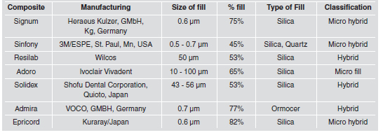
The aim of the study was to evaluate the effect of immersion media on the color stability of seven different composite resins (Solidex - Shofu, Resilab - Wilcos, Signum – Heraeus, Epricord - Tokuyama, Adoro – IvoclarVivadent, Admira - Voco and Sinfony - 3M ESPE). Their characteristics are listed in Table 1.
Table 1. Composite Resin used in this study.
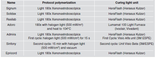
All resin specimens were prepared in cylindrical Teflon molds 10 mm in diameter and 2 mm thick. The molds were placed on a glass plate covered with a transparent mylar strip to get a flat surface. The molds were covered with another transparent strip which was pressed flush under a top glass plate. Each specimen was irradiated according to composition and manufacturer’s recommendations using tungsten-halogen lights box, specific for each composite (Table 2 and 3).
Table 2. Commercial name, manufacturer, composition and lot of resins used in the study.
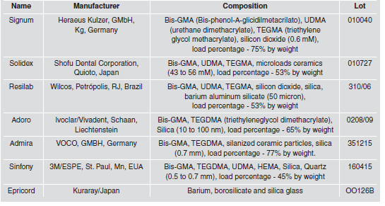
Table 3. Protocol polymerization of composite resin and light curing units used in this study.
Immediately after light activation, specimens were removed from the Teflon molds, finished and polished with 600-grit sandpaper disks. The finishing and polishing were done with the Sof-lex System – 3M ESPE and each disk was used intermittently for 15 seconds at a low speed. Fifteen specimens of each resin were chosen randomly and immersed in distilled water as control at 37°C for 24 hours. Following the first immersion cycle, all specimens were removed, rinsed under tap water, and blot-dried before color measurement. After initial measurement, the composite resin specimens were randomized into 3 groups (n=5) according to immersion media: coffee, cola beverage and water (control solution). The coffee solution was prepared by using 25 g of powder for 250 ml of water, standardized and changed every day.
The beverage used was Coca-Cola®; the pH of the cola beverage was measured using pH indicator paper (around 2.5) and also changed every day. The color of the specimens was measured with a spectrophotometer (Vita Easyshade, Vita Zahnfabrik, Bad S.ckingen, Germany). Colorimetric measurements of the specimens were performed on a white background according to the CIELab color scale, recording the L*, a*, and b* values. Three readings were performed and a mean value for the L*, a*, and b* values was obtained for each specimen. Color measurements were repeated after one week of immersion in the water control and in the two staining solutions. One examiner performed all the recordings of the L*, a*, and b* values. ΔE’ values were calculated using Hunter’s equation, ΔE’=[(ΔL*)2+(Δa*)2+(Δb*)2]1/2.
Statistical Analysis
The software SPSS® for Windows V.12.0 was used for statistical analysis. Descriptive statistics were shown as Mean±SD. Discoloration results of the specimens immersed in water, beverage and coffee for one week were analyzed using two-way repeated- measures ANOVA and Tukey’s comparisons post hoc test at 95% level of significance.
RESULTS
On the basis of these data, the hypothesis set as the premise of this study should be accepted. The staining ability of the resin composite materials is related to the type of staining agent. The considerations relative to these statements are presented below. According to the ANOVA two way test, the staining agents and their interaction with composite showed significant results (p=.00001) (Table 4). Mean values and SDs of color changes and group differences (ΔE) of micro hybrid resin composite restorative materials are listed in Table 5.
Table 4. Two-way ANOVA results for resin composite restorative materials and different staining agents.
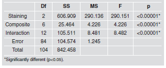
Table 5. Mean and SD of color changes and differences between groups after Tukey Comparisons Test of Delta for interaction staining/composite.
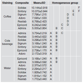
Tables 6 and 7 show the mean and SD for the two variables studied, type of composite and staining, and Fig. 1 and 2 show the lower influence of coffee on composites Admira and Epricord. For the microhybrid composite material groups, the lowest color differences (ΔE*) were observed in Admira and Resilab. When the beverage and coffee groups were compared, they both showed a higher color difference than the water group. The highest color change in micro-hybrid composites was observed in the Group Solidex (Table 5). When comparing the staining solutions, water, cola and coffee showed the highest mean ΔE* values and were significantly different for some of the resin composite materials tested. When comparing the seven different restorative materials, the cola solution/Admira group showed significantly less color change than the other materials tested. The greatest color difference in this study was observed in the Solidex and Sinfony material groups.
Table 6. Mean and SD of color changes and differences between groups after Tukey Comparisons Test of Delta for composite variable.
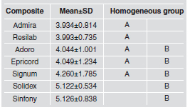
Table 7. Mean and SD of color changes and differences between groups after Tukey Comparisons Test of Delta for staining variable.
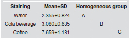
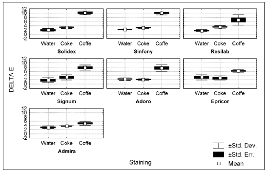
Fig. 1: Graphic Staining X ΔE.
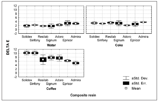
Fig. 2: Composite resin X ΔE.
DISCUSSION
Color stability has previously been studied in vitro for a variety of esthetic restorative materials10. Discoloration can be evaluated visually or by instrumental techniques11. The system for measuring chromaticity was chosen to record color differences because it is well suited to the determination of small color differences12. Colorimeters are capable of detecting color differences below the threshold of visual perception11. The value of ΔE* represents relative color changes that an observer might report for the materials after immersion or between time periods. Thus ΔE* is more meaningful than the individual L*, a*, b* values13. Discoloration can be evaluated with various instruments. Since instrument measurements eliminate the subjective interpretation of visual color comparison, spectrophotometers and colorimeters have been used for measuring color change in dental materials. When differences in color matching between human-eye and colorimetric assessment were compared, color difference was perceptible when ΔE values were greater than three10. The differences in optical properties due to water exposure might be explained by the material’s composition and the way that it is affected by environmental conditions. It is known from previous studies that resin-based composites allow water penetration into the matrix or filler-matrix interface. The presence of microcracks in the resin matrix as a result of swelling and plasticizing effects, along with interfacial gaps created between the filler and resin matrix allow stain penetration and discoloration of the restoration14. As it is assumed that the resin component is a source of discoloration, a high volume fraction of the resin would be expected to correlate with a high prospect for discoloration15. Low staining susceptibility is generally related to a low water absorption rate, and hydrophobic resins or low resin content in the composition of the restorative material. Traditional dimethacrylates form cross-linked networks with unreacted pendant methacrylates that serve as plasticizers; this plasticization imparts a more open structure to the polymers, which facilitate the absorption of dye. In addition, the increase in dye concentration means has been attributed to the porosity of some glass particles of the filler2. We demonstrated that the effect of the staining solutions was different for various materials. These results are in agreement with previous studies6,7,16. Previous studies testing longer staining periods have reported that after 7 days or longer immersion, materials had visually perceptible color changes7,13,15. Therefore the drinking habits of the patients must be considered when choosing restorative resin-based materials, especially at the front of the mouth.
During color measurement, both the actual color of the surface and the lighting condition under which the surface is measured will affect the measured color5. In the present study, a standard illuminant against a white background was used. As color difference evaluation was the focus of this study, the choice of illuminant was not important. When color is measured with an instrument that has a small window for both illumination and collection of light, a considerable fraction of the light entering the specimen is probably lost17. To minimize the edge loss effect, the diameter of the specimens( 10 mm) prepared in this study was greater than the aperture size of the instrument(3 mm × 8 mm). The degree of staining of indirect composite resin in this study is related to the adsorption and penetration of dyes in the organic matrix of resin because of the compatibility of the polymer phase with the yellow dye present in coffee, since the composite resin with higher degree of staining was immersed in soluble coffee solution (Table 7). Intense staining can be noted after the seven days of staining (Table 5), due to the process of absorption of coloring agents that occurs in the resins. Changes were continuously increasing, although it was not the objective of this study. Comparing these seven types of resin in relation to intrinsic stability and surface resistance to staining, Admira showed the smallest change (ΔE = 3.934 ± 0.814) and Sinfony the least influence of dye (ΔE = 5.126 ± 0.838). All values obtained by spectrophotometric system were higher than those clinically acceptable (ΔE> 3.3)
The degree of polarity of dyes determines their degree of penetration into the resin. Less polar dyes, such as coffee, can penetrate easily into the polymer matrix, while more polar dyes, such as wine, only impregnate the surface of the material20Another possible explanation for the degree of staining is the relationship between pH of the dye solutions and the composite resin. One previous study showed that acid dyes (pH 3-6) attacked the surface of dental materials and caused some changes in the surface integrity, softening the matrix and causing loss of structural ions such as Ca2 +, Al3 +, Sr2 +, Ba2 +, P3 + e Si2 + 23. However, another study examined the influence of pH on the sorption behavior and solubility of dental material and showed greater solubility in materials immersed in solution with pH 4 and 6, than with pH 8. It was observed that although the cola solution had a low pH, which could damage the surface integrity of the dental materials, it did not produce as much discoloration as coffee and tea. This may have been because the absence of caramel color in cola solution causes color changes ranging from palest yellow to deepest brown3. The according to Bagheri et al.18 and Ertas et al.19 the lack of yellow colorant in cola resulted in less discoloration than that caused by coffee. The sorption of water can rapidly degrade the composite, plasticizing the resin, hydrolyzing the silane and causing micro cracks. The micro cracks allow stain penetration and discoloration. A hydrophilic material has a higher degree of water sorption and discoloration20. In the present study, the specimens immersed in soluble coffee showed higher adsorption of dyes, which induced higher color instability compared to the other groups. This agrees with several previous studies, although the staining changes might be different in vivo because the period of absorption is shorter and the food is always new, whereas in this study, the solutions were changed every 24 hours to keep them fresh.
CONCLUSION
Within the limitations of this study, it could be concluded that coffee had a higher impact on the staining of the composite resin surface than cola, and that the composite resins Admira and Resilab showed the lowest discoloration means after immersion in staining solutions for seven days.
1. Puckett AD, Fitchie JG, Kirk PC, Gamblin J. Direct composite restorative materials. Dent Clin North Am 2007;51: 659-675. [ Links ]
2. Mundim FM, Garcia Lda F, Pires-de-Souza FdeC. Effect of staining solution and repolishing on color stability of direct composites. J Appl Oral Sci 2010;18:249-54. [ Links ]
3. Catelan A, Briso ALF. Sundfelf RH; Goiato MC, Santos PH. Color stability of sealed composite resin restorative materials after ultraviolet artificial aging and immersion in staining solution. J Prosthet Dent 2011;105:236-241. [ Links ]
4. Begum TS, Kocak A, Esra A. Effect of five staining solutions on the colourstabilityof two acrylics and three composite resins based provisional restorations. Eur J Prosthodont Restor Dent 2006;14:2-6. [ Links ]
5. Omata Y, Uno S, Nakaoki Y, Tanaka T, Sano H, Yoshida S, et al. Staining of hybrid composites with coffee, oolong tea, or red wine. Dent Mater J 2006;25:125-131. [ Links ]
6. Guler AU, Yilmaz F, Kulunk T, Guler E, Kurt S. Effects of different drinks on stainabilityofresin composite provisional restorative materials. J Prosthet Dent 2005;94:118-124. [ Links ]
7. Villalta P, Lu H, Okte Z, Garcia-Godoy F,Powers JM. Effects of staining and bleaching on color change of dental composite resins. J Prosthet Dent 2006;95:137-142. [ Links ]
8. Haselton DR, Diaz-Arnold AM, Dawson DV. Color stability of provisional crown and fixed partial denture resins. J Prosthet Dent 2005;93:70-75. [ Links ]
9. Okubo SR, Kanawati A, Richards MW, Childress S. Evaluation of visual and instrument shade matching. J Prosthet Dent 1998; 80:642-648. [ Links ]
10. Lee YK, Lim BS, Kim CW. Influence of illuminating and viewing aperture size on the color of dental resin composites. Dent Mater 2004;20:116-123. [ Links ]
11. Yap AU, Sim CP, Loh WL, Teo JH. Human-eye versus computerized color matching. Oper Dent 1999;24:358-363. [ Links ]
12. Khokhar ZA, Razzoog ME, Yaman P. Color stability of restorative resins. Quintessence Int 1991;22:733-737. [ Links ]
13. Yannikakis SA, Zissis AJ, Polyzois GL, Caroni C. Color stability of provisional resin restorative materials. J Prosthet Dent 1998; 80:533-539. [ Links ]
14. Bagheri R, Burrow MF, Tyas M. Influence of food simulating solution and surface finish on susceptibility to staining of aesthetic restorative materials. J Dent 2005;33:389-398. [ Links ]
15. Inokoshi S, Burrow MF, Kataumi M, Yamada T, Takatsu T. Opacity and color changes of tooth-colored restorative materials. Oper Dent 1996;21:73-80. [ Links ]
16. Turkun LS, Leblebicioglu EA. Stain retention and surface characteristics of posterior composites polished by one-step systems. Am J Dent 2006;19:343-347. [ Links ]
17. Van der Burgt TP, ten Bosch JJ, Borsboom PC, Kortsmit WJ. A comparison of new and conventional methods for quantification of tooth color. J Prosthet Dent 1990;63:155-162. [ Links ]
18. Watts DC, Bertenshaw BW, Jugdev JS. pH and time-dependence of surface degradation in a compomer biomaterial. J Dent Res 1995;74: 912. [ Links ]
19. Ertas E, Guler AU, Yucel AC, Koprulu H, Guler E. Color stability of resin composites after immersion in different drinks. Dent Mater J 2006;25:371-376 [ Links ]
20. Ortengren U, Andersson F, Elgh U, Terselius B, Karlsson,S. Influence of pH and storage time on the sorption and solubility behavior of three composite resin materials. J Dent 2001;29:35-41 [ Links ]

