


 Inglés (pdf)
 Articulo en XML
 Referencias del artículo
 Enviar articulo por email
 Citado por SciELO
  Similares en
SciELO
Similares en
SciELO 
 Permalink
INTRODUCTION
The pediatric patient shows certain anatomophysi ological characteristics that favor the development of respiratory complications, including narrowing of the airways, low functional residual capacity and nondevelopment of collateral ventilation1. Hospi talization in the PICU (Pediatric Intensive Care Unit) and specially the use of mechanical ventila tion (MV) add several factors that somewhat favor their appearance2,3. Also, analgesia and sedation used during ventilatory support have a relevant role, because they alter the protective mechanisms of the airways4. On the other hand, complications derived from the patients’ stay at the PICU, as, for example, acquired muscle weakness and loss of mobility, may cause retention of secretions and subsequent development of atelectasis, worsening the situation even more5.
Respiratory physiotherapy (RP) is a set of techniques that contribute to mucociliary clear ance, favoring the removal of secretions retained in patients with difficulty in managing secretions.6 Among these techniques, there is the group of “high frequency” techniques, which are the ones that generate high-frequency, low-volume oscillations during the expiratory phase and can be produced both in an active and a passive manner. These techniques can be used manually, through passive devices such as the Flutter® or Acapella®, or mechanically, with the high-frequency chest compressor (Vest®), the high-frequency chest oscillator (The Hayek oscil lator®) and intrapulmonary percussive ventila tion (IPV)7. The latter is a mechanical bronchial hygiene technique (MBHT) where a high-pass, pulsatile flow is combined with low tidal volumes delivered at high frequencies. This causes a transpulmonary positive pressure gradient that favors alveolar recruitment and secretion clear ance through an increase in the expiratory flow8 (Figure 1). In general, we can say there are two IPV devices available on the market. Devices used in hospitals are pneumatic and can incor porate high concentrations of oxygen (O2); some are even combined with aerosol delivery and con tinuous airway pressure (CPAP)9 whereas home devices are electric and only deliver IPV without any other additional resources. The frequency range used by these devices is 1.7-5 Hz, with pressures from 10 cmH2O up to 40 cmH2O; and they are usually used in sessions that may last between 15 and 20 minutes10,11. They can be used with a nasal or oronasal mask, a mouthpiece, or directly connected to the artificial airways with or without mechanical ventilation. the greatest advantage of the IPV in pediatric patients is capacity to achieve excellent coupling with spon taneous ventilation; also, it doesn’t need patient cooperation and has very good tolerance7,12,13.
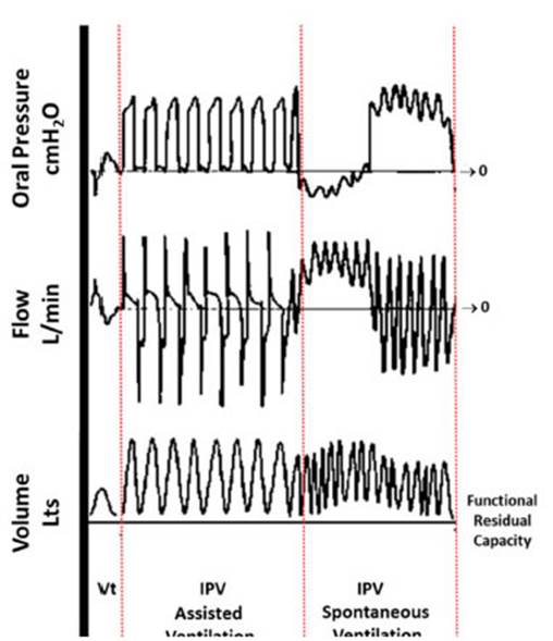
Figure 1 Graphic scheme of flow, volume and pressure curves during inspiratory and expiratory times with the use of IPV. Pressures are illustrated as measured in the mouth.
Several authors showed the efficacy of the IPV in different populations of pediatric patients14,15 and explained that the IPV is as effective as conventional bronchial hygiene techniques in patients with cystic fibrosis16. However, in pub lications related to the use of IPV in the PICU, we can see the lack of evidence in this field. The only randomized clinical trial (RCT) is the study in which it was proven that the IPV is a safe treatment and is effective for the resolution of atelectasis in pediatric patients on mechanical ventilation17.
Despite the fact that evidence in favor of us ing RP in the PICU is controversial in certain scenarios, there are studies that support the use of IPV in critical pediatric patients18,19. However, the number of worldwide publications about this topic so far is low and we couldn’t find any stud ies conducted in Latin America12,17,20. Therefore, the primary objective of our study was to describe the characteristics of the population in whom we used a home IPV device as MBHT in the PICU. Secondary we will describe the methodology for using this device and its results.
MATERIALS AND METHODS
This study is a prospective, observational and descriptive case series conducted in the Juan P. Garrahan pediatric hospital in the Autonomous City of Buenos Aires, Argen tina, in the period between August 1st, 2019 and December 31st, 2019.
The study included patients younger than 18 years on ventilatory support who received at least one session of IPV in the intensive care unit.
For this study, an electronic record charts was designed with private access from the mobile devices of the research ers. These were used to record demographic variables, such as gender, age in months, weight, and also variables related to the initial diagnosis, as for example, the pres ence of some kind of complex chronic condition (CCC), the reason for using ventilatory support, whether it was type 1 or type 2 acute respiratory failure (ARF) and the type of initial ventilatory support21. Also, IPV-related variables were recorded: indications for IPV (atelectasis, hypersecre tion, hypoxemia), treatment duration, parameters of each session, I/E (inspiration/expiration) ratio, pressure range, amount of cycles (number of fractions of time within each session), in-line or independent use, and type of ventilatory support required by the patient at the beginning of each session (invasive mechanical ventilation, IMV; non-invasive mechanical ventilation, NIMV; high-flow nasal cannula, HFNC, or extracorporeal membrane oxygenation, ECMO). The use of additional O2 during treatment, complications, and clinical and result parameters were recorded during each session.
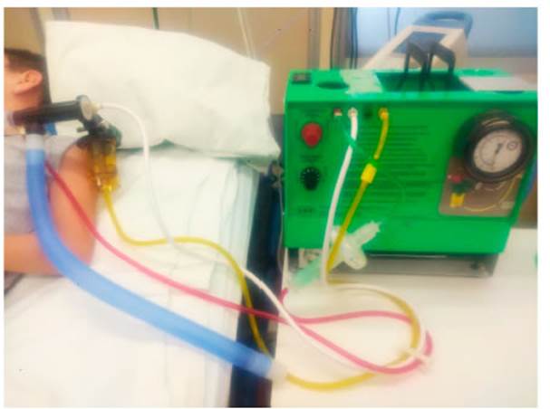
The following home IPV devices were used for this study: The Impulsator ® from Percussionair (Sandpoint, Idaho, United States) (Figure 2) with Phasitron ® circuits (Sandpoint, Idaho, United States). The parameters used at the discretion of the physiotherapist according to the objective and tolerance of the patient were: frequency of 90 cycles/min, 180 cycles/min and 250 cycles/min, I/E ratio of 1:1, 2:1 and 3:1 and a maximum pressure range of 10-40 cmH2O22,23. In the cases where the device was used in-line, it was set in assist pressure-controlled ventilation (PA/C-CMV) mode, with a positive end-expiratory pressure (PEEP) of more than zero24. At the beginning of each ses sion, the patient was placed lying on his/her back with a head elevation of 30°. In patients with arterial O2 saturation decrease below 88%, the corresponding cycle was suspended and continuation of treatment was reconsidered according to clinical tolerance. If the event was repeated, the session was suspended. As regards the duration of each session, we considered a maximum of 20 minutes. In patients with atelectasis, the frequency of the sessions was at least two per day, with one or two sessions in the 8-16 h period and another one, according to the criterion of the treating physiotherapist, in the 16-24 h period, during on-call time. In patients with hypersecretion, the IPV was adapted to their bronchial hygiene plan, according to the criterion of the health professional.
Due to their asymmetrical distribution, continuous variables are expressed as medians and interquartile ranges (IQR), and categorical variables are expressed as frequen cies and percentages. For the data analysis, we used the IBM SPSS® Macintosh, version 25.0 (IBM Corp., Armonk, NY, USA) statistical package.
Given the fact that it is an observational study, informed consent wasn’t required. During the whole process, data were kept confidential and the identity of the patients was preserved through numerical codification.
RESULTS
Eighteen patients were included in the study; 33% of the patients were female, with a median of age of 13.5 (4.75 - 54) months and a median of weight of 10 (5-13.5) kg (Table 1).
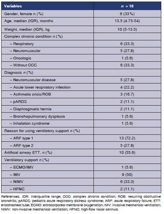
The 66.7% of the patients had a CCC at study entry; the most common diagnosis was neuromus cular disease (27.8%), and the reason for requiring ventilatory support was type 1 acute respiratory failure (ARF) in 72.5% of the cases. A total of 48 IPV sessions were carried out (Table 2).
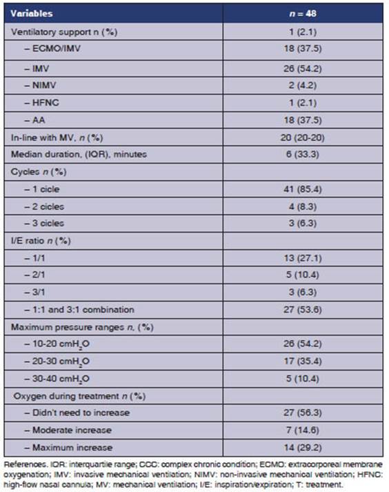
The 38.9% of the patients received one session; another 38.9% received two, and the remaining 22.2% received three or more. The main reason for IPV use was the atelectasis diagnosis (83.3%); other reasons were hypersecretion and hypox emia. The treatment was carried out both in patients who were on IMV (55.6%) and patients with non-invasive support (44.4%), whether it was NIMV or HFNC. In 58.4% of the sessions, the patients were receiving some sort of non-invasive ventilatory support. In 22.9% of the cases, the treatment was carried out in-line with the ventilator. In the patient who was on ECMO, the IPV wasn’t in-line.
The median duration of each session was 20 minutes. In 85.4% of the cases, it was done in only one cycle without interruption. With regard to the way of using the IPV, it was set with a 3:1 I/E ratio in 59.9% of the sessions; in 89.6% the maximum pressure was lower than 30 cmH2O; and in 43.7% of the sessions, it was necessary to increase the O2 during the treatment. 80% of the patients who had sessions with a maximum pressure range of 30-40 cmH2O were on IMV with endotracheal tube.
The 53.3% of patients showed radiographic resolution of atelectasis, where 75% only required between one and two sessions to resolve it. The saturation/fraction of inspired O2 ratio (SpO2/FiO2) improved in 14.6% of the sessions.
In 6.3% of the sessions, it wasn’t possible to complete the prescribed time due to desatura tion. However, two out of three patients who had presented this complication resolved the atelectasis.
DISCUSSION
This study allowed us to describe the population receiving IPV within the environment of our in stitution, a reference center for Latin America. It also showed us that it is possible to use home IPV devices in the PICU, since they are effective with only a small number of sessions for the resolution of atelectasis.
The demographic characteristics of our study were similar to those of Morgan et al. mostly male patients, 63% of patients with a median of age of 2 years, and a median of weight of 14 kg20. The characteristics were also similar to the Deakins group, which reported a median age of 3.1 years17.
The CCC of neurological origin is a character istic that limits the patient’s capacity to remove secretions effectively. In our study, the group of patients with this condition required IPV just like all the patients included in the study of Bidwala et al., who had fewer annual infections and used less antibiotics and steroids13.
Birkrant et al. proposed that more research should be done for the purpose of finding which are the diseases that would benefit most from the use of this technique and describing more complica tions during its implementation; accordingly, we provided a variety of diagnoses, healthy patients and patients with CCC, patients with invasive and non-invasive support and some minor complica tions25.
The major problem with respiratory physiother apy in the pediatric population is mainly the lack of cooperation and irritability to certain stimuli. For that reason, the IPV could be presented as a good alternative to other techniques in young chil dren, since it doesn’t require patient cooperation. Individual setup of percussion and frequency is generally well-tolerated, because it doesn’t require coordination with the patient; on the contrary, it is adjusted to the patient’s respiratory rate17,22,25. It is even a good bronchial hygiene alternative in patients of less than one year with gastroesopha geal reflux, as shown in the study of Lievens et al26.
In most studies, there wasn’t any adverse event12,13,16,17,27. Only Morgan and Homnick de scribed an episode of pneumothorax and one mild hemoptysis, respectively20,28. Unlike those cases, we had three mild desaturation levels immedi ately solved after treatment discontinuation. Some laboratory studies allow us to understand that IPV would represent a low risk for barotrauma, in terms of the pressure levels used, especially in pediatric patients on IMV29. Certain authors observed the pressure drop phenomenon: with similar maximum inspiratory pressure compared to the PA/C-CMV mode, the mean airway pressure in an IPV system is much lower. This could be due to the shape of the pressure curve and the effect of percussions on pulmonary geometry, which could produce a drop in the alveolar pressure30,31. Another relevant piece of information observed by Smallwood is that the percentage of pressure reduction is inversely proportional to the size of the tube, which reaches up to 60% in the 3.0 tube. This observation favors its use in the neonatal and pediatric populations29.
According to Deakins, the in-line use of IPV in patients who received invasive mechanical ventila tion is safe and effective for the resolution of atel ectasis, just like it was observed in our population. However, we believe our case series study provides information mainly relating to the diversity of this MBHT in patients with different clinical situations and ventilatory needs17.
In the study of Rivera Cano et al., this technique was used as NIMV in patients with bronchiolitis who didn’t respond to CPAP, but it wasn’t used as a MBHT. Regarding the implementation of IPV in patients on ECMO, we could only find one case report of a pediatric patient with Bordetella Pertussis who received IPV to resolve a massive at electasis, with good treatment response32,33. There are studies where IPV was used in patients with ARDS or ECMO as invasive ventilatory support, but not as a MBHT, which is the object of study of this article34-37.
Pediatric publications regarding the use of IPV as a MBHT in critical patients are limited but promising, since they show the fact that it is a safe and effective option for the resolution of atelectasis. The resolution rate of our study was 53.3%. The Deakins study used an atelectasis score that emphasizes a large improvement in the IPV group compared to the group receiving conventional respiratory physiotherapy, with a median of 3.1 days versus 6.2 days that the control group took for the resolution3,17. In our study, the group of patients who resolved the at electasis did it in a shorter period of time, since they required only a median of two sessions. This difference could be due to the fact that the duration of the sessions in our study was longer (10 minutes versus 20 minutes), and that, at the moment of the Deakins et al. publication, there was no awareness about the way of optimizing the setup of the IPV device24. Yen Ha et al. could have provided additional data on this regard, but, the methodology of radiographic diagnosis (all the patients underwent X-rays on day 2 and then on day 5) may have limited the findings, since there could have been patients that immediately resolved the atelectasis but still continued with the treatment for 5 days12. The IPV is an effective MBHT that helps clear secretions and takes little time to resolve atelectasis. Like other authors, we believe this characteristic could be relevant due to the reduction in physiotherapy treatment time and healthcare costs13,17,19.
With respect to the duration of the IPV ses sions, we know there are two ways of setting up the device that vary between 10 and 20 minutes. The reason for the shorter duration could be as sociated with two situations: one is the fact that there are some IPV devices such as the MetaNeb® System (Hillrom Services, Amsterdam, the Neth erlands) that have a 10-minute timer13,20; and the other situation is that the session ends when the nebulizer runs out of physiological solution, which lasts for approximately 10-15 minutes; that is the criterion for ending the session14,17. Both in the work of Yen Ha et al. and our work, the usage trend was between 15 and 20, as suggested by the Guides published by the creator of this technology, Dr. Forrest Bird10,12.
Some authors describe the setup of the IPV, but generally not in a specific way. In his in vitro study about the effects generated in the flow and pressure by the different ways of setting up the device, Toussaint showed that the expiratory flow increased with increasing I:E ratio. This coincides with the I:E setup trend chosen by physiotherapists in our hospital for 59.9% of the sessions12,13,22.
A common characteristic in pediatric publica tions about IPV was the small number of patients. The study with the largest number of patients was the retrospective descriptive analysis of Morgan et al., with 59 patients on IMV and in-line use of IPV20; in the RCT of Deakins et al., a total of 12 patients were admitted (5 in the control group and 7 in the intervention group); Campbell Reardon et al. included 18 neuromuscular patients; Yen Ha et al., 6 patients with atelectasis and breathing difficulty; and Bidiwala et al., 8 tracheostomized chronically critically ill patients12-14,17. Our study with more than 18 patients provides information about the versatility of the technique and treat ment of atelectasis, since it includes more cases than the rest of the PICU publications on this topic.
LIMITATIONS
The fact that we didn’t use a score for the diagnosis of atelectasis could be considered a bias; however, we chose not to use it because it is underused in our work environment so, instead, we decided to use common radiographic techniques for this purpose. Oxygenation through arterial gases could be an outcome variable to consider in terms of the effi cacy of the technique; we used SpO2/FiO2 to avoid unnecessary invasive processes. The measurement of the volume of secretions was dismissed, because there isn’t any standardized method for this. Our work doesn’t allow the generalization of the re sults, but due to the characteristics of the patients of our hospital, it wouldn’t be completely wrong to consider it as a useful tool to optimize secretion management in patients who need it and to treat atelectasis in the PICU.
CONCLUSION
This study describes the population in which IPV is population in which IPV is implemented in the context of an institution in Argentina, which is positioned as a benchmark in Latin America. It also presents a tool that could be useful for the resolution of atelectasis within a short period of time, optimizing its use in healthcare practice.

