


Servicios Personalizados
Revista

Articulo
 texto en
  Español (pdf)
 Articulo en XML
 Referencias del artículo
 Enviar articulo por email
Indicadores
-
 Citado por SciELO
Links relacionados
-
 Similares en
SciELO
Similares en
SciELO
Compartir

 Permalink
Revista argentina de cirugía
versión On-line ISSN 2250-639X
Rev. argent. cir. vol.113 no.1 Cap. Fed. abr. 2021
http://dx.doi.org/10.25132/raac.v113.n1.1472.ei
Articles
Mucinous adenocarcinoma of the gallbladder originated from intracholecystic papillary neoplasm
1 División de Cirugía General del Hospital Dr. Cosme Argerich. Buenos Aires. Argentina.
Gallbladder cancer is the most common malignancy of the biliary tract, ranking sixth among gastrointestinal cancers, and is characterized by its aggressive and poor prognosis. Complete resection with chemotherapy is the only possibility of cure and long-term survival depending on tumor stage.
In the pancreatobiliary tract, tumors composed of preinvasive neoplastic cells that form clinically detectable (≥ 1.0 cm) masses are classified as intraductal papillary neoplasms (BilIN) in the bile ducts and as intraductal papillary mucinous neoplasms or intraductal tubulopapillary neoplasms (ITPNs) in the pancreas. Similar lesions have been described in the gallbladder and have been classified under a unified category of intracholecystic papillary neoplasm (ICPN)1. According to the 2010 WHO classification, this lesion is characterized by an exophytic intramucosal mass that measures > 1.0 cm and is composed of preinvasive neoplastic (dysplastic) cells forming a compact lesion distinct from the neighboring mucosa5.
Intracholecystic papillary neoplasm with invasive mucinous adenocarcinoma and signet ring cells is a variety of gallbladder cancer. The aim of this article is to describe a case of a rare gallbladder cancer with specific histology, and the treatment performed.
We report the case of a 73-year old female patient who sought medical care due to pain in the right hypochondriac region radiating to the epigastrium and associated with nausea, vomiting and loss of weight within the past two months. The laboratory tests were normal, including liver panel and tumor markers. The ultrasound showed distended gallbladder, focal anterior wall thickening at the fundus with multiple gallstones inside (Fig. 1). Magnetic resonance cholangiopancreatography and computed tomography scan revealed a dilated common bile duct, with distal tapering of the ampulla of Vater, multiple endoluminal filling defects all along its length and heterogeneous wall thickening in the gallbladder fundus (Fig. 2).
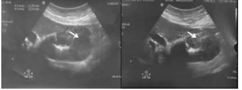
Figure 1 Ultrasound. The gallbladder is distended, with gallstones and focal thickening of the fundus (arrow)

Figure 2 A. Computed tomography scan showing wall thickening of the gall bladder fundus (black arrow). B. Magnetic resonance cholangiopancreatography showing a dilated common bile duct of 10.3 mm, multiple endoluminal filling defects (white arrow), gallbladder (arrowhead).
The patient underwent exploratory laparoscopy. There were no metastases in the liver or peritoneum. The gallbladder was resected through open surgery and a frozen section biopsy was performed (mucinous papillary neoplasm). The procedure included resection of liver segments IVb/V, bile duct, clearance of the hepatic pedicle lymph nodes and Roux-en-Y hepaticojejunostomy.
The histopathology report made the diagnosis of intracholecystic papillary neoplasm with associated invasive mucinous adenocarcinoma with signet ring cells involving the liver (Fig. 3). The resection margins were clear, and 9/9 lymph nodes were involved. The final tumor staging was pT3: cancer with tumor extending directly into the liver bed; pN2: cancer with metastases in 4 lymph nodes or greater.

Figure 3 A and B. Intracystic papillary neoplasm with invasive mucinous adenocarcinoma, (white arrow). The liver (white arrowhead) and gallbladder wall are held with a clamp in A. C. Microscopic image showing associated signet ring cell carcinoma (black arrow).
The patient evolved with favorable postoperative outcome and is currently receiving adjuvant therapy (chemotherapy).
Gallbladder cancer is rare, and several histological subtypes have been described: adenocarcinoma, papillary adenocarcinoma, mucinous adenocarcinoma, signet ring cell carcinoma, adenosquamous carcinoma, neuroendocrine carcinoma, small cell carcinoma and undifferentiated carcinoma2. Yet, only 2.5% correspond to mucinous adenocarcinomas and 0.82% are associated with signet ring cell carcinoma as in our patient. Mean age of presentation is 65 years independently of sex and 87% are diagnosed as pT3 stage with unusually aggressive behavior. Survival at 3 years is 1% compared with 39% of other gallbladder adenocarcinomas3.
Intracystic papillary neoplasia (ICPN) is a histologically non-invasive lesion, which produces mucus secretion responsible for the inflammation of the gallbladder and resembles cholecystitis in imaging tests. It can also appear as a tumor in the gallbladder fundus4. Although the preoperative diagnosis made with imaging tests may not be conclusive, it can provide a diagnostic suspicion, helping to ensure timely and appropriate treatment as in the present case5.
In our patient, the tumor size was > 6 cm and extended directly into the liver and perimuscular connective tissue without compromising the serous layer, as mentioned by Sugawara et al. and Manterla et al.1,5. Most cases are papillary tumors are 50% are invasive, with an immunohistochemical profile that is different from conventional gallbladder cancer due to its expression of MUC1 and MUC23.
Intracholecystic papillary neoplasm with invasive mucinous adenocarcinoma and signet ring cells is a rare variety of gallbladder cancer with symptoms mimicking cholecystitis. Survival and prognosis are worse than in other types of gallbladder cancer despite surgery and chemotherapy.
Acknowledgments
We are especially grateful to Department of Anatomical Pathology, Diagnostic Imaging and Oncology Services of Hospital Argerich, who contributed to the diagnosis and treatment of the patient.
REFERENCES
1. Manterola C, Bellolio E, Otzen T, Duque G. Neoplasia papilar intra colecística. Reporte de un caso y revisión de la evidencia existen te. Int J Morphol. 2018; 36(4):1485--9. [ Links ]
2. Pina LN y cols. Carcinoma incidental de vesícula biliar en un Hospi tal Universitario. Acta GastroenLatam. 2017; 47(3):190-93. [ Links ]
3. Dursun N, et al. Mucinous Carcinomas of the Gallbladder Clini copathologic Analysis of 15 Cases Identified in 606 Carcinomas. Arch Pathol Lab Med. 2012; 136(11):1347-58. [ Links ]
4. Mizobuchi N, et al. Three Cases of Intracystic Papillary Neoplasm of Gallbladder. Abdom Radiol. 2018; 43(7): 1535-9. [ Links ]
5. Sugawara S, et al. 2018. A Case of Mucinous Cystic Neoplasm of the Gallbladder. Clinical Journal of Gastroenterology. 2018; 11(5): 42832. https://doi.org/10.1007/s12328-018-0850-8. [ Links ]
Received: January 16, 2020; Accepted: June 23, 2020
 Este es un artículo publicado en acceso abierto bajo una licencia Creative Commons
