


 Inglês (pdf)
 Artigo em XML
 Referências do artigo
 Enviar este artigo por email
 Citado por SciELO
  Similares em
SciELO
Similares em
SciELO 
 Permalink
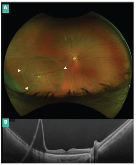
A 37-year old male had recently noted a nasal scotoma in his right eye (OD). His best corrected visual acuity was 20/20 in both eyes. Fundus examination of the OD revea led a thin, smooth, immobile, dome-shaped and bullous elevation of the retina at the inferotemporal quadrant, which showed to be a retinoschisis extending from the periphery to the vascular arcade at the same quadrant, just less than 2-disc diameters from the fovea (Fig. 1A, white arrowheads mark its edges). Left eye was normal. There is no relevant personal or family medical history. Imaging with an integrated ultra-wide field swept sour ce optical coherence tomography device in OD (blue line in Figure 1A shows area cut out by OCT) was performed and confirmed the diagnosis of acquired bullous retinos chisis showing the intraretinal cleavage at the external plexiform layer from the macular periphery (Fig. 1B). No active behavior was indicated. He remained stable after a 6-month follow-up.
Acquired retinoschisis may be unilateral or bilateral. It can rarely be bullous or expand to the macula, as in our case. Conversely, X-linked congenital retinoschisis predo minantly affects the fovea, presenting as a cystic change in the posterior pole but, in nearly half cases, it associates to acquired peripheral retinoschisis.

