


Serviços Personalizados
Journal

Artigo
 Artigo em XML
 Referências do artigo
 Enviar este artigo por email
Indicadores
-
 Citado por SciELO
Links relacionados
-
 Similares em
SciELO
Similares em
SciELO
Compartilhar

 Permalink
Acta Odontológica Latinoamericana
versão On-line ISSN 1852-4834
Acta odontol. latinoam. vol.22 no.1 Buenos Aires abr. 2009
ARTÍCULOS ORIGINALES
Influence of nutritional state on radiographic variables in upper canine teeth
Luis F. Wuscovi1, Hugo N. Aragón2, María E. Gordillo3, María E. López3
1 Department of Radiology,
2 X-Ray Room and
3 Department of Biological Chemistry. School of Dentistry. Universidad Nacional Tucumán.
CORRESPONDENCE Od. Luis Fernando Wuscovi Cat. Radiologia - Fac. Odontologia- UNT Av. Benjamin Araoz 800 (4000) S. M. de Tucuman, Argentina Fax: 54 381 4227589 Phone: 54 381 4107308 E-mail: luisfernandowuscovi@hotmail.com Home Phone: 54 381 4284039
ABSTRACT
Permanent upper canine tooth impaction is related to both genetic causes and nutritional and local causes. Based on radiographic studies of children, Haavikko, 1974, calculated the median of the degree of calcification for each tooth according to age. The aim of our study was to establish associations between certain radiographic variables with respect to upper canine tooth buds and nutritional state in a population of schoolchildren. Thirty-three children of chronological ages 3 to 9 years from a population of rural schoolchildren took part in the study. The children were classified into three groups: malnourished, control and overweight. For the radiographic study we used a systematized technique using DSJ 70kV radiographic equipment with 8 mA and Insight intraoral film (E F speed; Eastman Kodak, Rochester, NY). The images obtained from the 66 radiographic studies were scanned. The program Image tool for Windows was used to measure: a) distance from the canine cusp to the intermaxillary suture, b) outer angle formed between the axis of the canine tooth and the plane that cuts the intermaxillary suture perpendicularly, c) degree of calcification d) presence of supernumerary teeth. The values of these variables were analyzed statistically with the Kruskal- Wallis test of the SPSS program. The results were: a) For the distance variable there is no significant difference between the malnourished and control groups (p>0.05), although there are differences (p<0.001) between the control and overweight groups; b) There is no significant difference between nutritional states for the angle variable; c) For the ratio between chronological age and dental age calculated according to degree of calcification, significant differences were found between the malnourished and overweight groups (p< 0.01), while the control group showed no difference regarding Haavikko’s values (p>0.05). Both the distance of the canine tooth bud and its degree of calcification vary in patients with nutritional disorders, therefore it would be useful to conduct a radiographic study at school age to detect tooth anomalies and reduce their consequences.
Key words: Radiographic studies; Nutritional state; Canine tooth bud.
RESUMEN
Influencia del estado nutricional sobre variables radiográficas de canino superior
La retención de los caninos superiores permanentes se encuentra asociada tanto a causas genéticas como nutricionales y locales. A partir de estudios radiográficos en niños, Haavikko, 1974, calculo la mediana del grado de calcificación de cada pieza dentaria según la edad. El objetivo de este estudio fue establecer asociaciones entre ciertas variables radiográficas respecto al germen de los caninos superiores y al estado nutricional de una población escolar. Se trabajo con 33 niños de edad cronológica entre 3 y 9 años de una población escolar rural. Se clasificaron los niños en los grupos desnutrido, control y sobrepeso. Para el estudio radiográfico se aplico una técnica sistematizada utilizando un equipo radiografico DSJ de 70kV con 8 mA y películas intraorales Insight (E F speed; Eastman Kodak, Rochester, NY). De los 66 estudios radiográficos obtenidos se escanearon las imagenes. Con el programa Image tool para Windows se midieron: a) distancia de cuspide de canino a sutura intermaxilar b) Angulo externo formado entre el eje del canino y el plano que corta la sutura intermaxilar en forma perpendicular c) grado de calcificación d) presencia de dientes supernumerarios. Los valores de estas variables se analizaron estadísticamente con el test de Kruskal-Wallis del programa SPSS. Los resultados fueron: a) Para la variable distancia no existen diferencias estadísticamente significativas entre los grupos desnutrido y control (p>0,05), aunque existen diferencias (p<0,001) entre los grupos control y sobrepeso; b) No existen diferencias significativas entre los estados nutricionales para la variable angulo; c) Para la relación entre la edad cronológica con respecto a la edad dental calculada a traves del grado de calcificación, se observaron diferencias significativas entre los grupos desnutrido y sobrepeso (p< 0,01), en tanto que el grupo control no presento diferencias con respecto a los valores de Haavikko (p>0,05). Tanto la distancia del germen del canino como su grado de calcificación varían en pacientes con trastornos nutricionales por lo que seria de utilidad la aplicación de un estudio radiográfico a edad escolar para detectar anomalías dentarias y reducir sus consecuencias.
Palabras clave: Estudios radiograficos; Estado nutricional; Germen canino.
INTRODUCTION
Permanent upper canine teeth follow third molars in frequency of impaction, with an incidence of 0.8 to 2.8%1, 2, which has been found to be 2.2% for populations of Swedish schoolchildren3 and 3% for a Saudi population4. Frequency of impaction is three times higher in females than in males5. The causes of canine tooth impaction may be general or localized6. Generalized causes include endocrine deficiencies, febrile diseases and radiation, while localized causes are most often the case in impaction, and include such causes as discrepancy between arch length and tooth size, lengthy impaction or premature loss of the deciduous canine tooth, abnormal position of the tooth bud, defect of the alveolus or alveolar cleft, anchylosis, cysts or neoplastic formations, root dilaceration, iatrogenic causes7, traumas8 and idiopathic causes. Some authors report genetic9, 10 or family factors11. Palatal impaction of the permanent upper canine tooth appears in several generations of one family, and is related to congenital missing teeth or microdontia, especially of the upper lateral incisors12. Another factor that affects tooth development is nutrition, as delayed development of tooth buds has been reported in patients with malnutrition and patients with low birth weight, with associated malocclusion13.
Statistical determination of nutritional levels is based on three indicators: weight-age, which measures overall malnutrition; height-age, which reflects chronic malnutrition because shortness is the result of a lengthy lack of nutrients, and weight-height, which measures acute malnutrition14. The prevalent type in northern Argentina is height-age deficit. Measurements of dental arches in children chronologically aged 4 to 8 years with different nutritional states were significantly larger in females with normal nutritional state than in females with chronic malnutrition; no difference was found among males15. Tooth embryology and formation is a continuous process of development that runs from the sixth week of prenatal life to about 20 years of age. Of all teeth, the permanent upper canine has the longest development period; it emigrates from the side of the pyriform fossa to its final destination in the occlusion position16. During the course of its development, the crown is closely related to the root of the upper lateral incisor, and it has been determined statistically that there is a relationship between palatal displacement of the permanent upper canine tooth and absence or reduction in crown width of the adjacent lateral canine tooth17.
Different methods, all of which provide similar results, can be used to estimate dental age according to the radiographically observed degree of development of the permanent tooth buds18. In this study we used the method proposed by Haavikko, 1974, which was obtained from Scandinavian populations19, because there are no original tables for tooth development in the population of northern Argentina. The radiographic factors that influence orthodontic decisions to expose or remove an impacted upper canine tooth have been evaluated. These factors are palatal position of the crown and angling towards the mid-line20. Some researchers suggest paralleling studies, where the horizontal angling is altered enabling determination of the position of the canine impacted in the occlusal plane towards the vestibular or palatal location. In images this is seen as a displacement of the canine towards mesial or distal positions. The combination of periapical studies with occlusal studies has also been mentioned21. During clinical practice, the first radiograph is often a panoramic radiograph, in which the image of the impacted canine teeth in palatal position is magnified with respect to canine teeth located in normal position. Computed tomography is the most sensitive, specific method22, 23. Other authors, using profile teleradiography, found that ectopic canine teeth are displaced further forward than normal teeth24.
Because of the multiple consequences of impacted canine teeth, such as external root resorption of neighboring teeth, mainly the upper lateral incisors25, crown resorption26 and the fact that they may turn into dentigerous cysts27, early diagnosis of this kind of anomaly is important The aim of this study was to establish associations between certain radiographic variables related to the upper canine tooth buds and the nutritional state of a population of schoolchildren in northern Argentina.
MATERIALS AND METHODS
The study included 33 children of chronological ages 3 to 9 years from a population of rural schoolchildren in Tucuman. Height and weight were recorded using precision scales with a height rod, which was calibrated prior to the start of the study. The operator underwent theoretical and practical training in nutritional anthropometry according to the recommendations of the World Health Organization27. Chronological ages were obtained by checking school records. Parents were asked for informed consent. According to the height-age and weight-age values assi gned in the Argentine Pediatrics Association guidelines for assessment of growth28 the children were classified into 3 nutritional groups: a) Malnourished: values beneath percentile 25; b) Normal: values between percentiles 25 and 75; c) Overweight: values above percentile 7529 (Lejarraga and Orfila, 1987).
Radiographic examination: A systematized radiographic technique was applied to both the right and the left permanent upper canine teeth sides. Wheeled 70kV DSJ 8 mA X-ray equipment and Insight No 2 intraoral film (E F speed; Eastman, Kodak, Rochester, NY) were used. The following procedure was applied: a) Head position: obtained when the Frankfurt plane that goes from the wing of the nose to the tragus (lateral view) and the bipupilar plane (front view) are parallel to the ground (Fig. 1). b) Position of the film: parallel to the occlusal plane, held in place by bite pressure, so that the greater axis is parallel to the intermaxillary suture going beyond the mid-line, while the shorter edge of the film, located in anterior position, should go beyond the incisor edges of the upper central incisors by 1 mm, in parallel form. c) Direction of the central ray: in antero posterior direction; the ray must be directed with a vertical angulation of 70o with respect to the plane of the film, and must be parallel to the mid-line, i.e. 0o horizontal angulation is applied, with a point of incidence at the level of the canine fossa (Fig. 2). d) Mean exposure time: 0.40 seconds. Radiographic material was processed manually, and patients received all the adequate radio-protection measures (law 17557).
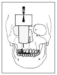
Fig. 1: Front view of the application of the systematized special technique.
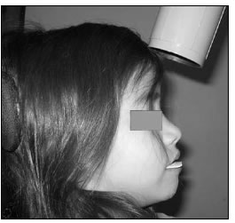
Fig. 2: Vertical angulation with respect to the horizontal plane. Profile view of the systematized special technique.
The 66 radiographic studies obtained were digitalized with a scanner (Genius, Color Page-HR3), and the Image Tool for Windows 3.00 was used to measure the following variables: a) distance from the upper canine cusp to the intermaxillary suture, expressed in mm (Fig. 3); b) external angle formed between the axis of the permanent upper canine and the plane cutting the intermaxillary suture perpendicularly (Fig. 4), expressed in degrees (o); c) calculation of dental age was determined by means of observation of the degree of calcification of the buds of the upper canine teeth in the radiographic images30, according to Haavikko, 1974 19. This method divides tooth development into 11 phases, 5 of which correspond to crown development, and 6 to root phases: Crypt or absence of calcification (0), initial calcification (C1), half crown development (Cr .), three quarters crown development (Cr .), complete crown development (Crc), initial root formation (Ri), quarter root length (R .), half root length (R .), three quarters root length (R .), full root length (Rc) and closed apex (Ac).
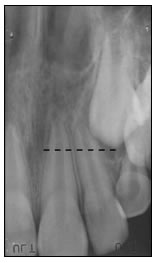
Fig. 3: Measurement of the distance from the canine cusp to the middle raphe in a digitalized radiographic image.
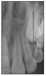
Fig. 4: Measurement of the external angle formed between the axis of the upper canine tooth bud and the perpen - dicular plane that cuts the in - ter maxillary suture.
Statistical analysis: The statistical analysis was done with the software SPSS version 11 (SPSS Inc., IL, USA) using the normal distribution tests (Kolmogorov- Smirnov and Shapiro-Wilk).
In order to determine whether there are differences between the external angle and distance variables with respect to the nutritional states, Kruskal-Wallis tests and Dunn multiple comparison tests were used. A paired T test was used to relate dental age according to Haavikko, 197419, to the corresponding chronological age for the groups with different nutritional states.
RESULTS
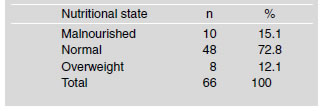
Regarding the three nutritional groups, it was found from the height-age and weight-age records that 15.1% of the children (n=10) were malnourished, 72.8% (n=48) were normal and 12.1% (n=8) were overweight (Table 1).
Table 1: Distribution of children according to nutritional state
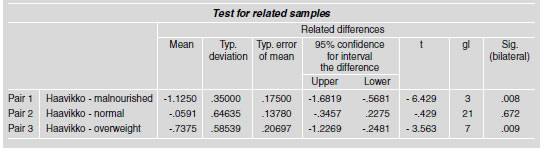
In the 66 radiographic studies conducted, the distance and angle variables for the three nutritional groups did not have a normal distribution (Fig. 5a and b, Table 2). For the distance variable, there were significant differences between the normal and overweight groups (p<0.05), but not so between the normal and malnourished groups (p>0.05). The angle variable showed no statistically significant difference among the three nutritional groups (p>0.05). Regarding the analysis of degree of calcification of permanent upper canine tooth buds, Table 3 shows the values for the children’s chronological age and age according to Haavikko, 1974, and the statistics obtained for the three nutritional groups. The analysis of dental age, obtained according to the degree of canine calcification in relation to chronological age, the malnourished and overweight groups (paired T test) showed significant differences (p<0.05). The age of the normal group with respect to Haavikko, 197419, showed no significant difference (p>0.05) (Table 4).
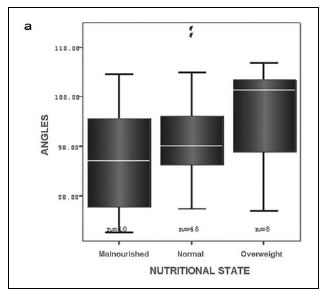
Fig. 5: Non-normal distribution of the data of the variables angle (a) and distance (b), according to nutritional condition as from the median.
Table 2: Normality test for the variables angle and distance according to nutritional state
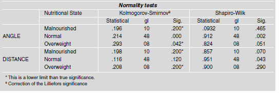
Table 3: Statistics of chronological age for each nutritional state and age calculated using de Haavikko, 1974 tables
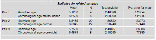
Table 4: T-test for related samples of the pairs formed between age of the nutritional groups and age calculated using Haavikko (1974) tables, according to degree of calcification of the permanent canine
DISCUSSION
Tucuman Province has one of the highest rates of child malnutrition (30%)28. These values do not include overweight children, whose condition is often the result of poor quality nutrition made up mainly of low-cost food containing many calories and low nutritional value. This study involves the odontological and radiographic clinical analysis of parameters which allow alterations in the speed of development of tooth buds to be detected and could contribute to early detection of a potential impacted upper canine tooth. Radiographically measured variables are identified which enable the detection of alterations in the speed of development of the upper tooth buds. These alterations are also associated to children’s nutritional state. Some authors believe that the best age for early diagnosis of future impacted upper canine teeth is from 8 to 10 years because at that age the canine tooth still has its eruptive potential. That would allow its position to be corrected by means of orthodontic treatment without traumatic procedures such as surgical exposure and traction. The removal of the temporary canine tooth is also suggested for patients whose arches do not lack space, in order to avoid potential impaction31, 32. Aesthetic and functional disorders, and even lateral incisor resorption, are thus avoided33.
The study of dental calcification is considered to be the best method for determining a patient’s biological age. Many methods have been developed to determine the degree of calcification of the permanent tooth buds based on radiographic images, some of which require the information of a quadrant, while others, such as Haavikko, 1974, and Nolla, 196034, suggest using a tooth or small group of teeth, thus reducing the time and cost involved in the study, which in turn enables dental age to be determined in cases where information on the whole quadrant is unavailable. This is why the Haavikko, 1974, method was chosen for this study, as well as because it has proved to be an effective estimator of chronological age in children of both genders under 10 years of age35. Since Tucuman Province has not published its own tables for standard weight and height for girls and boys, this study used as a reference the tables drawn up from populations in Buenos Aires and Cordoba Provinces and La Plata city, Argentina29, to calculate the normal nutritional state of the population.
The application of a systematized radiographic technique prevents factors not dependent on the clinical case from altering the values of the variables. As conventional radiographic equipment and standard film are used, the technique is inexpensive and can be applied by a dentist general practitioner. This study showed that both the distance of the upper canine tooth bud and its degree of calcification vary in patients with nutritional disorders, so it would be useful to conduct a systematic radiographic study at school age in order to detect anomalies in dental development and thus reduce their possible consequences.
ACKNOWLEDGMENTS
This study was partly funded by the Technical and Science Office, Research Council at Tucuman National University (J310).
1. Shah RM, Boyd MA, Vakil TK. Studies of permanent tooth anomalies in 7886 Canadian individuals. I: Impacted teeth. J Canad Dent Assoc 1978;44:262-264. [ Links ]
2. Grover PS, Lorton L. The incidence of unerupted permanent teeth and related clinical cases. Oral Surg, Oral Med, Oral Pathol and Endod 1985;59:420-425. [ Links ]
3. Thilander B, Myrberg N. The prevalence of malocclusion in Swedish school children. Scand J Dent Res 1973;81:12-21. [ Links ]
4. Zahrani AA. Impacted cuspids in a Saudi population: prevalence, etiology and complications. Egypt Dent J 1993; 39:367-374. [ Links ]
5. Ericson S, Kurol J. Early treatment of palatally erupting maxillary canines. Eur J Orthod 1988;10:283-295. [ Links ]
6. Bishara SE, Kommer DD, Mc Neil MH. Management of impacted canines. Am J Orthod 1976;80:173-190. [ Links ]
7. Bishara SE. Clinical management of impacted maxillary canines. Semin Orthod 1998;4:87-98. [ Links ]
8. Brin I, Solomon Y, Zilberman Y. Trauma as a possible etiologic factor in maxillary canine impaction. Am J Orthod Dentofacial Orthop 1993;104:132-137. [ Links ]
9. Pelsmaekers B, Loos R, Carels C, Derom C, Vlietinck R. The genetic contribution to the dental maturation. J Dent Res 1997;76:1337-1340. [ Links ]
10. Peck S, Peck L, Kataja M. The palatally displaced canine as a dental anomaly of genetic origin. Angle Orthod 1994;64:249-256. [ Links ]
11. Pirinen S, Arte S, Apajalahti S. Palatal displacement of canine is genetic and related to congenital absence of teeth. J Dent Res 1996;75:1742-1746. [ Links ]
12. Mossey P, Campbell HM, Luffingham JK. The palatal canine and the adjacent lateral incisor: a study of a west of Scotland population. Br J Orthod 1994;21:169-174. [ Links ]
13. Quinonez Ybarra ME, Rodriguez Calzadilla A, Gonzalez Cabrera B, Padilla Gonzalez CM. Morbilidad bucal. Su relacion con el estado nutricional en ninos de 2 a 5 anos de la consulta de nutricion del Hospital Pediatrico Docente de Centro Habana. Rev Cubana Estomatolol 2004; 41(1) http://www.imbiomed.com/1/1/articulos.php?id_revista=63&id_ejemplar=2860
14. World Health Organization, Physical Status: the use and interpretation of anthropometry. WHO Technical report series 845. Ginebra, 1995.
15. Moreno Rodriguez K, Meneses Lopez A, Morzan Valderrama E. Dimensiones de arcos dentarios en ninos de 4 a 8 anos de edad con diferente estado nutricional: Talara-Piura. Rev Estomatol Herediana 2004;14:18-21. [ Links ]
16. Stivaros N, Mandall NA. Radiographic factors affecting the management of impacted upper permanent canines. J. Orthod 2000;27:169-173. [ Links ]
17. Marinelli A, Nannelli P. Diagnostic evaluation of abnormally erupted maxillary canines. Minerva Stomatol 1999; 48:265-271. [ Links ]
18. Demirjian A, Goldstein H, Tanner JM. A new system of dental age assessment. Hum Biol 1973;45:211-227. [ Links ]
19. Haavikko K. Tooth formation age estimated on a few selected teeth. A simple method for clinical use. Proc Finn Dent Soc 1974;70:15-19. [ Links ]
20. Jacobs S.G. Localization of the unerupted maxillary canines: how to and when to. Am J Orthod Dentofacial Orthop 1999;115:314-322. [ Links ]
21. Armstrong C, Johnston C, Burden D, Stevenson M. Localizing ectopic maxillary canines-horizontal or vertical parallax. Eur J Orthod 2003;25:585-589. [ Links ]
22. Fox NA, Fletcher GA, Horner K. Localizing maxillary canines using dental panoramic tomography. Br Dent J 1995;179:416-420. [ Links ]
23. Gavel V, Dermaut L. The effect of tooth position on the image of unerupted canines on panoramic radiographs. Eur J Orthod 1999;21:551-560. [ Links ]
24. Mcsherry P, Richardson A. Ectopic eruption of the maxillary canine quantified in three dimensions on cephalometric radiographs between the ages of 5 and 15 years. Eur J Orthod 1999;21:41-48. [ Links ]
25. Filippi A. Reabsorcion de tejidos dentarios por terceros molares retenidos ectopicos. Quintessence 1995;8:73-77. [ Links ]
26. Azaz B, Shteyer A. Resorption of the crown in impacted maxillary canine. A clinical radiographic and histologic study. Int J Oral Surg 1978;7:167-171. [ Links ]
27. World Health Organization. Medicion del efecto nutricional de programas de suplementacion alimentaria a grupos vulnerables OMS Ginebra 1980. [ Links ]
28. Sociedad Argentina de Pediatria. Criterios de diagnostico y tratamiento. Crecimiento y desarrollo. Buenos Aires, 1997. [ Links ]
29. Lejarraga H, Orfila G. Estandares de peso y estatura para ninos y ninas argentinos desde el nacimiento hasta la madurez. Arch Argent Pediatr 1987;85:209-222. [ Links ]
30. Borman T, Solheim B, Magnusson, SI. Inter-examiner variation in the assessment of age-related factors in teeth. Int J Legal Med 1995;107:183-186. [ Links ]
31. Shapira Y, Kuftinec MM. Early diagnosis and interception of potential maxillary canine impaction. J Am Dent Assoc 1998;129:1450-1454. [ Links ]
32. Jacobs SG. Reducing the incidence of palatally impacted maxillary canines by extraction of deciduous canines: a useful preventive/interceptive orthodontic procedure. Case reports. Aust Dent J 1992;37:6-11. [ Links ]
33. Richardson G, Russell KA. A review of impacted permanent maxillary cuspids diagnosis and prevention. J Can Dent Assoc 2000;66:497-501. [ Links ]
34. Nolla C. The development of the permanent teeth. J Dent Child 1960;27:254-266. [ Links ]
35. Bolanos MV, Manrique MC, Bolanos MJ, Briones MT. Approaches to chronological age assessment based on dental calcification. Forensic Sci Int 2000;110:97-106. [ Links ]

