


Serviços Personalizados
Journal

Artigo
 Inglês (pdf)
 Artigo em XML
 Referências do artigo
 Enviar este artigo por email
Indicadores
-
 Citado por SciELO
Links relacionados
-
 Similares em
SciELO
Similares em
SciELO
Compartilhar

 Permalink
Acta Odontológica Latinoamericana
versão On-line ISSN 1852-4834
Acta odontol. latinoam. vol.24 no.1 Buenos Aires abr. 2011
ARTÍCULOS ORIGINALES
Influence of the hybrid layer thickness and resin tag length on microtensile bond strength
Vanessa Rahal1, André L.F. Briso1, Paulo H. dos Santos2, Maria L.M.M. Sundefeld3, Renato H. Sundfeld1
1 Department of Restorative Dentistry, Araçatuba Dental School, São Paulo State University, Araçatuba, São Paulo, Brazil.
2 Department of Dental Material and Prosthodontics, Araçatuba Dental School, São Paulo State University, Araçatuba, São Paulo, Brazil.
3 Department of Community and Preventive Dentistry, Araçatuba Dental School, São Paulo State University, Araçatuba, São Paulo, Brazil.
CORRESPONDENCE Vanessa Rahal, Discipline of Restorative Dentistry – Sao Paulo State University. Rua Jose Bonifacio 1193, Sao Paulo, Sao Paulo, Brazil. 16015-050. Email: vanessarahal@hotmail.com Phone/fax: +55 18 36363346.
ABSTRACT
The purpose of this study was to evaluate the correlation between the hybrid layer thickness/resin tag length and the microtensile bond strength of conventional two-step adhesive system, when applied to healthy dentinal tissue. After performing the restorative adhesive procedures and tooth extractions, ten specimens were sectioned in the mesiodistal direction. One section was used for microscopic analysis of the resin tag lengths and the hybrid layer thickness, while the other was used for the microtensile bond strength test (0.5 mm/min). The fractured surface was classified according to the fracture pattern, under a stereoscopic microscope at 40× magnification. Data obtained were submitted to analysis using one-way ANOVA and Pearson’s Correlation test (α=0.05). The means corresponding to the hybrid layer thickness, resin tag lengths and the microtensile test were 2.68 μm, 6.43 μm and 16.23 MPa, respectively. There was no correlation between the means of the values obtained for the microtensile test, and those presented by the hybrid layer (r2=0.40, p>0.05) and resin tags (r2=0.21, p>0.05). The microtensile bond strength of the conventional twostep adhesive system Adper Single Bond 2 did not depend on the thickness of the hybrid layer and length of resin tags.
Key words: Dental adhesive; Dental materials; Microtensile bond strength; Microscopy.
RESUMO
Influência da espessura da camada híbrida e comprimento dos tags resinosos na resistência adesiva a microtração
A proposta deste estudo foi avaliar a correlacao entre a espessura da camada hibrida e comprimento dos tags com a resistencia adesiva a microtracao de um adesivo convencional de dois passos, quando aplicado em dentina higida. Depois da realizacao dos procedimentos restauradores e extracoes dentarias, dez especimes foram seccionados no sentido mesio-distal. Uma das seccoes foi utilizada para analise microscopica dos tags resinosos e da espessura da camada hibrida, e a outra seccao foi utilizada para o teste de microtracao (0,5 mm/min). A superficie fraturada foi classificada de acordo com o padrao de fratura, sob lupa estereoscopica em 40×. Os dados obtidos foram submetidos ao ANOVA e teste de correlacao de Pearson (α=0,05). As medias correspondentes a espessura da camada hibrida, comprimento dos tags e do teste de microtracao foram 2,68 μm, 6,43 μm and 16,23 MPa, respectivamente. Nao houve correlacao entre as medias dos valores obtidos para o teste de microtracao e camada hibrida (r2=0,40, p>0,05) e microtracao e tags resinosos (r2=0,21, p>0,05). O teste de microtracao do adesivo convencional de dois passos Adper Single Bond 2 nao apresentou correlacao com a espessura da camada hibrida e o comprimento dos tags resinosos.
Palavras-chave: Adesivos dentarios; Materiais dentarios; Resistencia a microtracao; Microscopia.
INTRODUCTION
The clinical success of adhesive restorations in dentin is directly related to the physical and mechanical properties of the adhesive system to be used, which contribute to the bond strength of adhesive materials in the face of the action of masticatory forces1-3.
Similarly, it has also been observed and reported4,5 that the quality of the bond interface formed in dentin is variable and dependent on various factors, among them, the hybrid layer and resin tags. In line with these surveys, the quality of the bond interface formed in clinical6,7 and laboratory8 studies is known with regard to the most diverse adhesive systems, such as those that advocate total acid etching prior to their application4,9,10 or self-etching systems11,12. One of the ways to observe the effectiveness of the dentinal bond of an adhesive material is by means of common optical microscopy, which provides consistent information in larger areas of the adhesive interface, according to the reports of some authors4,8. On the other hand, the bond effectiveness may also be obtained by the microtensile test, which enables greater distribution of forces on the bond interface, resulting in reliable tensile strength values13-15. With the use of this method, various studies have shown that bond strength depends, among other factors, on the quality of the dentinal tissue16 and the adhesive system used 4,17; however, few have discussed the correlation between the hybrid layer thickness4,8, resin tag lengths and tensile strength. It should be pointed out that at the moment, although the available literature18,19 emphasizes that dentinal acid etching enables high bond strength values to be obtained, the correlation of bond strength with the capacity of the bonding agent to penetrate into dentinal tissue and formation of the hybrid layer, still requires further explanations.
Therefore, the proposal of this study was to analyze the correlation of the length of resin tags and the thickness of the hybrid layer, with the microtensile bond strength of a conventional two-step adhesive system, Adper Single Bond 2, when applied to dentinal tissue in vivo. The null hypothesis tested was that the bond strength was not influenced by the hybrid layer thickness and resin tag length.
MATERIALS AND METHODS
Specimen Preparation and Restorative Procedures
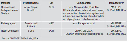
After the project had been submitted to and approved by the appropriate Research Ethics Committee, ten third molars indicated for extraction were selected. After dental prophylaxis, local anesthesia of the operative field was performed and wide Class I restorations were made, involving practically the entire occlusal surface, to a depth of 4 mm. The diamond bur 1094 (KG Sorensen Industria e Comercio Ltda., Barueri, Sao Paulo, Brazil) was mounted in a high-speed handpiece, and used under water and air cooling (MS 350 - Dabi-Atlante, Ribeirao Preto, Sao Paulo, Brazil). Before performing the restorative procedures, whenever possible the teeth received rubber dam isolation, however, for those that presented difficult access, due to the patient’s buccal anatomy, careful relative isolation of the operative field was performed. A conventional two-step adhesive system Adper Single Bond 2 (3M/ESPE, St Paul, MN, USA) was used, as well as resin composite Z-250 (3M/ESPE, St Paul, MN, USA), whose specifications are described in Table 1.
Table 1: Materials used in the study.
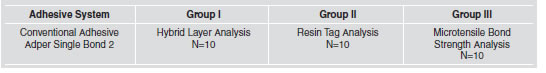
The ten teeth were divided into three groups according to the type of analysis to be performed on dentinal tissue, considering the following factors: resin tags, hybrid layer, and microtensile bond strength (Table 2).
Table 2: Study Groups.
The cavities were etched with 35% phosphoric acid Scotch Etchand (3M/ESPE, St Paul, MN, USA) for 15 seconds and after this period, abundantly washed for 20 seconds. The etched surface was kept humid, the excess water being removed with a small cotton wool ball. After this, the Adper Single Bond 2 adhesive (3M/ESPE, St Paul, MN, USA) was applied to the tooth surface with a microbrush (Microbrush Inc., Grafton, MA, USA), according to the manufacturer’s instructions. After the application of the adhesive, smooth air jets were applied for 30 seconds to allow the solvents to evaporate. After this, the adhesive was light-activated for 20 seconds at 450 mW/cm2 with a light polymerization appliance (Ultralux Lens – Dabi Atlante, Ribeirao Preto, Sao Paulo, Brazil), regularly checked with a radiometer (100, KERR Corporation, Orange, CA, USA). At this time, the cavities received the resin composite Z-250 (shade A2) (3M ESPE Dental Products, St Paul, MN, USA) inserted in oblique increments. Each layer was light-polymerized for 40 seconds. After polymerizing the layers of resin composite, all the restorations were finished with diamond burs of the Gold Series No.1190F (K.G. Sorensen Industria e Comercio Ltda., Barueri, Sao Paulo, Brazil), followed by the use of Enhance siliconized burs (Dentsply - Industria e Comercio Ltda., Petropolis, Rio de Janeiro, Brazil) for finishing resin restorations.
Immediately after this, the restored teeth were extracted, taking the necessary care not to traumatize the restored area. Afterwards, the teeth were cleaned and sectioned in the mesio-distal direction, using a diamond disk under constant irrigation, in a metallographic cutter, ISOMET 1000 (Buehler, Lake Bluff, IL, USA), thus obtaining vestibular and lingual sections.
Optical Microscopy Analysis
One of the dental sections was used for optical microscopy, and was decalcified in a solution containing equal proportions of 50% formic acid and 20% sodium citrate. Complete decalcification of each specimen was verified by taking radiographs8. After decalcification, the restorations were carefully removed, and the remaining organic material was carefully embedded in paraffin. The specimens were sectioned in the longitudinal direction of the clinical crown to a thickness of six micrometers, and sequentially mounted on glass slides. Fifteen slides of each specimen were randomly selected to be stained with the Brown & Bren method. The best histological sections of each slide were analyzed using Axiophot optical mi - cros copy (ZEISS DSM-940 A, Oberkochen, Baden-Wurttemberg, Germany) at 1000× magnification, with a micrometric eyepiece 100×/1.30 (Fig. 1). The measurements of the hybrid layer and resin tags were taken after meticulous analysis of the entire extent of the histological section using the computer program AxioVision Software Real (Version 4.3; Carl Zeiss MicroImaging Inc., Thornwood, NY, USA). For each factor under analysis, three measurements were obtained in each section, in which the resin tags analyzed and measured belonged to the same region of the hybrid layer analyzed and measured. Consequently, for each selected slide, the thickness of the hybrid layer and length of the resin tags corresponded to the mean of the three measurements taken. Therefore, for each specimen, fifteen measurements were obtained for the factor hybrid layer, as well as for the factor resin tags.
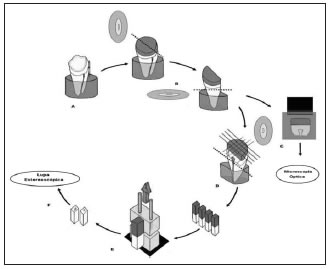
Fig. 1: Methodology used in the study. (A) Restorative Procedure. (B) Cutting the teeth into two sections. (C) Optic Microscopy Analysis (D) Transversal sections to obtain stick-shaped specimens for the microtensile test. (E) Microtensile test (F) Analysis of the fracture surface by stereoscopic microscope.
Microtensile Tests
The other tooth section was used for the microtensile bond strength test. For this purpose, a metallographic cutter ISOMET 1000 (Buehler, Lake Bluff, IL, USA) was used, under water cooling at a speed of 800 rpm and static load of 80 grams, to allow sections to be performed in planes transverse to the dentin bond/resin composite interface, in order to obtain stick-shaped specimens. These specimens were fixed to the Universal Test Machine Instron Model 4411 (Instron Inc., Canton, MA, USA), with cyanoacrylate adhesive (Super Bonder - Henkel Ltda., Itapevi, Sao Paulo, Brazil) and tested with a 50N load cell at a speed of 0.5 mm/min until they ruptured.
Fracture Pattern Analysis
The fracture pattern was analyzed under a stereoscopic microscope Stemi SV 11 (Carl Zeiss Company - DSM-940 A, Oberkochen, Baden- Wurttemberg, Germany) at 40× magnification. To reveal the fracture area of each specimen more clearly, the revealing solution proposed by Ohkubo et al., in 198220, was applied in the fractured region, which stained the resin-free dental structure. To analyze the fracture patterns the criteria proposed by Figueiredo Reis et al. 21 were used (Table 3).
Table 3: Criteria for Fracture Pattern Analysis (Figueiredo Reis et al., 200321 with modifications).

Data Analysis
The results obtained for hybrid layer thickness and tags length and also microtensile bond strength were submitted to analysis of variance (ANOVA) and to Pearson’s Correlation test, at a level of significance of α=0.05, to verify whether there was correlation between the resin tag lengths, hybrid layer thickness and the bond strength of the studied adhesive system. Moreover, percentage comparison between the fracture patterns was performed to illustrate and elucidate the failures occurred in the specimens used in micotensile bond test.
RESULTS
Analysis by optical microscopy showed uniform resin tags (Fig. 2) with a mean length of 6.43 μm and mean hybrid layer thickness of 2.68 μm, which was uniform and present throughout the full extent of the sections; the microtensile test showed a mean value of 16.23 MPa. Application of Pearson’s correlation test, between the means of the factors microtensile bond strength and hybrid layer thickness (r2=0.40, p>0.05) and between those of microtensile bond strength and resin tag length (r2=0.21, p>0.05), showed absence of statistically significant correlation between these factors (Fig. 3 and 4).

Fig. 2: Resin tags analyzed by common optical microscopy (Group II – E1) (1000× magnification).
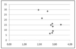
Fig. 3: Correlation Test between hybrid layer and microtensile bond strength for the adhesive system Adper Single Bond 2.
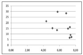
Fig. 4: Correlation Test between resin tags and microtensile bond strength for the adhesive system Adper Single Bond 2.
Fracture Pattern Analysis
In the tested specimens 42% adhesive fracture pattern, 32% mixed fractures and 19% cohesive fractures in resin and 7% in dentin were observed (Fig. 5). This means that most of the failures occurred in the adhesive interface.
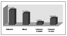
Fig. 5: Fracture Patterns for the adhesive system Adper Single Bond 2.
DISCUSSION
The analysis and measurement of the resin tags and hybrid layer by optical microscopy4,8, in addition to being a less expensive methodology, enabled a larger area of the bond interface to be analyzed, which is not usual in scanning electron microscopy22-24. During analysis of the histological sections, the contrast provided between the dentinal tissue and the resin structures intensely stained by the Brown & Brenn staining process, enables exceptionally good microscopic visualization of the desired resin structures4,8.
Certainly, the resin tags and hybrid layers found in this study were products of the complete removal of the dentinal smear layer and smear plugs, thanks to the previous etching with 35% phosphoric acid, which promoted exposure of the dentinal tissue collagen fibers 11,25,26, and infiltration of the adhesive system. After in vivo application of the conventional twostep adhesive system, mean hybrid layer thickness, resin tag length and microtensile bond strength values of 2.68 μm, 6.43 μm and 16.23 MPa, respectively were obtained, (Table 1); these values were not all that considerable, when compared with those obtained in vitro studies4,8,27. One of the reasons that could have contributed to the results obtained appears to be directly related to the internal pressure of the intratubular liquid, which probably made it difficult for the adhesive system to penetrate to a greater extent. Some studies have stated that the presence of intratubular pressure favors the obtainment of lower bond strength values than those obtained in situations in which there is no pulp pressure 28.
We may also consider the viscosity of the adhesive system used which, due to containing load particles in its composition, could possibly have contributed to its penetrating into the dentinal tubules to a lesser extent. According to the reports of Can Say et al., in 2006 29, the concentration of load particles in the dentinal tubule openings could reduce penetration of the adhesive system into them. The non-correlation found between the length of resin tags and microtensile bond strength of the conventional adhesive system Adper Single Bond 2, was reported in a previous study30 and can be explained by the direction of the application of forces during the microtensile test, which could reduce the mechanical strength of the adhesive31. The hybrid layer and resin tags formed in humid dentin have hydrophyllic molecules of low molecular weight in their composition (HEMA), which migrate to the deeper regions, mixing with the dentinal fluids and solvents of the adhesive system itself, thereby being weakly polymerized32; this may explain the adhesive fractures found during the fracture pattern analysis for this adhesive system. In a recent study, Anchieta et al.4, for this same adhesive system, observed in vitro the existence of correlation between the hybrid layer thickness and microtensile bond strength, however, the results observed in the present study showed no significant correlation between these factors; possibly due to the fact that this adhesive system had been applied in vivo, which would result in the occurrence of difficulty of complete evaporation of the solvent, humidity of the oral cavity and dentinal fluids, the low degree of conversion due to the presence of water, the inability to hybridize all of the collagen exposed during acid etching, which would form a region with lower fracture strength, or even due to the occurrence of hydrolytic degradation of the adhesive material, commonly described in the literature 33-36. This degradation has been proved by tests for nanoleakage at the bond interface37,38.
The null hypothesis was not rejected, since the microtensile bond strength of the conventional twostep adhesive system Adper Single Bond 2 did not correlate with the thickness of the hybrid layer and length of resin tags. According to the results observed in this study, it could be concluded that the microtensile bond strength of the conventional two-step adhesive system Adper Single Bond 2 did not depend on the thickness of the hybrid layer and length of resin tags.
1. Koshiro K, Sidhu SK, Inoue S, Ikeda T, Sano H. New concept of resin-dentin interfacial adhesion: the nano interaction zone. J Biomed Mater Res B Appl Biomater 2006;77:401- 408. [ Links ]
2. Masri M, Pilo R, Brosh T. The influence of convergence angle and dentin micromorphology on shear bond strength of adhesive resin luting cement. J Adhes Dent 2008;10: 277-284. [ Links ]
3. Wang Y, Spencer P. Hybridization efficiency of the adhesive/ dentin interface with wet bonding. J Dent Res 2003;82: 141-145. [ Links ]
4. Anchieta RB, Oliveira FG, Sundfeld RH, Rahal V, de Alexandre RS, Marquezini Jr L, Sundefeld MLMM. Evaluation of the correlation of hybrid layer and resin tags with the microtensile Bond strength of a conventional adhesive system applied on intact dentin tissue. Compend Contin Educ Dent. Forthcoming 2011. [ Links ]
5. Anchieta RB, Rocha EP, Ching-Chang KO, Sundfeld RH, Martin Junior M, Archangelo CM. Localized mechanics of dentin self-etching adhesive system. J Appl Oral Sci 2007;15:321-326. [ Links ]
6. Peumans M, Kanumilli P, De Munck J, Van Landuyt K, Lambrechts P, Van Meerbeek B. Clinical effectiveness of contemporary adhesives: a systematic review of current clinical trials. Dent Mater 2005;21:864-881. [ Links ]
7. Ritter AV, Swift EJ Jr, Heymann HO, Sturdevant JR, Wilder AD Jr. An eight-year clinical evaluation of filled and unfilled one-bottle dental adhesives. J Am Dent Assoc 2009;140: 28-37. [ Links ]
8. Sundfeld RH, Valentino TA, de Alexandre RS, Briso AL, Sundefeld ML. Hybrid layer thickness and resin tag length of a self-etching adhesive bonded to sound dentin. J Dent 2005;33:675-681. [ Links ]
9. Fusayama T. Posterior adhesive composite resin: a historic review. J Prosthet Dent 1990;64:534-538. [ Links ]
10. Nakabayashi N, Saimi Y. Bonding to intact dentin. J Dent Res 1996;75:1706-1715. [ Links ]
11. Reis A, Grandi V, Carlotto L, Bortoli G, Patzlaff R, Rodrigues Accorinte Mde L, Dourado Loguercio A. Effect of smear layer thickness and acidity of self-etching solutions on early and long-term bond strength to dentin. J Dent 2005;33:549-559. [ Links ]
12. Van Meerbeek B, Kanumilli P, De Munck J, Van Landuyt K, Lambrechts P, Peumans M. A randomized controlled study evaluating the effectiveness of a two-step self-etch adhesive with and without selective phosphoric-acid etching of enamel. Dent Mater 2005;21:375-383. [ Links ]
13. Pashley DH, Sano H, Ciucchi B, Yoshiyama M, Carvalho RM. Adhesion testing of dentin bonding agents: a review. Dent Mater 1995;11:117-125. [ Links ]
14. Sano H, Shono T, Sonoda H, Takatsu T, Ciucchi B, Carvalho R, et al. Relationship between surface area for adhesion and tensile bond strength evaluation of a micro-tensile bond test. Dent Mater 1994;10:236-290. [ Links ]
15. Sonoda H, Banerjee A, Sherriff M, Tagami J, Watson TF. An in vitro investigation of microtensile bond strengths of two dentine adhesives to caries-affected dentin. J Dent 2005;33:335-342. [ Links ]
16. Nakajima M, Sano H, Burrow MF, Tagami J, Yoshiyama M, Ebisu S, et al. Tensile bond strength and SEM evaluation of caries-affected dentin using dentin adhesives. J Dent Res 1995;74:1679-1688. [ Links ]
17. Yoshiyama M, Urayama A, Kimochi T, Matsuo T, Pashley DH. Comparison of conventional vs self-etching adhesive bonds to caries-affected dentin. Oper Dent 2000;25:163-169. [ Links ]
18. Barkmeier WW, Cooley RL. Laboratory evaluation of adhesive systems. Oper Dent 1992;Sup 5:50-61. [ Links ]
19. Triolo PT, Swift Jr EJ, Barkmeier WW. Shear bond strengths of composite to dentin using six dental adhesive systems. Oper Dent 1995;20:46-50. [ Links ]
20. Ohkubo N, Iwata S, Chikada K, Kuriyama S, Narita M, Ishikawa T, et al. A retention comparison of two sealants. Bull Tokyo Dent Coll 1982;23:201-219. [ Links ]
21. Figueiredo Reis A, Giannini M, Ambrosano GM, Chan DCN. The effects of filling techniques and a low-viscosity composite liner on bond strength to class II cavities. J Dent 2003;31:59-66. [ Links ]
22. Hsu KW, Marshall SJ, Pinzon LM, Watanabe L, Saiz E, Marshall GW. SEM evaluation of resin-carious dentin interfaces formed by two dentin adhesive systems. Dent Mater 2008;24:880-887. [ Links ]
23. Osorio R, Toledano M, de Leonardi G, Tay F. Microleakage and interfacial morphology of self-etching adhesives in class V resin composite restorations. J Biomed Mater Res B Appl Biomater 2003;66:399-409. [ Links ]
24. Radovic I, Vulicevic ZR, Garcia-Godoy F. Morphological evaluation of 2- and 1-step self-etching system interfaces with dentin. Oper Dent 2006;31:710-718. [ Links ]
25. Nakabayashi N, Ashizawa M, Nakamura M. Identification of a resin-dentin hybrid layer in vital human dentin created in vivo: durable bonding to vital dentin. Quintessence Int 1992;23:135-141. [ Links ]
26. Van Meerbeek B, De Munck J, Yoshida Y, Inoue S, Vargas M, Vijay P, Van Landuyt K, Lambrechts P, Vanherle G. Buonocore memorial lecture. Adhesion to enamel and dentin: current status and future challenges. Oper Dent 2003;28:215-235. [ Links ]
27. Arrais CAG, Giannini M. Morphology and thickness of the diffusion of resin through demineralized or unconditioned dentinal matrix. Braz Oral Res 2002;16:115-120. [ Links ]
28. Campos EA, Correr GM, Leonardi DP, Barato-Filho F, Gonzaga CC, Zielak JC. Chlorhexidine diminishes the loss of bond strength over time under simulated pulpal pressure and thermo-mechanical stressing. J Dent 2009; 37:108-114. [ Links ]
29. Can Say E, Nakajima M, Senawongse P, Soyman M, Ozer F, Ogata M, et al. Microtensile bond strength of a filled vs unfilled adhesive to dentin using self-etch and total-etch technique. J Dent 2006;34:283-291. [ Links ]
30. Lohbauer U, Nikolaenko SA, Petschelt A, Frankenberger R. Resin tags do not contribute to dentin adhesion in selfetching adhesives. J Adhes Dent 2008;10:97-103. [ Links ]
31. Misra A, Spencer P, Marangos O, Wang Y, Katz JL. Micromechanical analysis of dentin/adhesive interface by the finite element method. J Biomed Mater Res B Appl Biomater 2004;70:56-65. [ Links ]
32. Spencer P, Wang Y. Adhesive phase separation at the dentin interface under wet bonding conditions. J Biomed Mater Res 2002;62:447-456. [ Links ]
33. Ding PG, Wolff D, Pioch T, Staehle HJ, Dannewitz B. Relationship between microtensile bond strength and nanoleakage at the composite-dentin interface. Dent Mater 2009;25:135-141. [ Links ]
34. Sidhu SK, Omata Y, Tanaka T, Koshiro K, Spreafico D, Semeraro S, et al. Bonding characteristics of newly developed all-in-one adhesives. J Biomed Mater Res B Appl Biomater 2007;80:297-303. [ Links ]
35. Van Landuyt KL, Kanumilli P, De Munck J, Peumans M, Lambrechts P, Van Meerbeek B. Bond strength of a mild self-etch adhesive with and without prior acid-etching. J Dent 2006;34:77-85. [ Links ]
36. Yoshida Y, Nagakane K, Fukuda R, Nakayama Y, Okazaki M, Shintani H, et al. Comparative study on adhesive performance of functional monomers. J Dent Res 2004;83: 454-458. [ Links ]
37. Li H, Burrow MF, Tyas MJ. The effect of load cycling on the nanoleakage of dentin bonding systems. Dent Mater 2002;18:111-119. [ Links ]
38. Sano H. Microtensile testing, nanoleakage, and biodegradation of resin-dentin bonds. J Dent Res 2006;85:11-14. [ Links ]

