


Serviços Personalizados
Journal

Artigo
 texto em
  Espanhol (pdf)
 Artigo em XML
 Referências do artigo
 Enviar este artigo por email
Indicadores
-
 Citado por SciELO
Links relacionados
-
 Similares em
SciELO
Similares em
SciELO
Compartilhar

 Permalink
Revista argentina de cirugía
versão On-line ISSN 2250-639X
Rev. argent. cir. vol.113 no.1 Cap. Fed. abr. 2021
http://dx.doi.org/10.25132/raac.v113.n1.1493.ei
Articles
Laparoendoscopic resection of a tumor of the gastroesophageal junction
1 Unidad de Esófago, Hospital Universitario Fundación Favaloro. Buenos Aires. Argentina
Surgical resection of gastrointestinal stromal tumors (GIST) with negative margins is the standard of care for these tumors1. Atypical wedge resection through laparoscopy for gastric GIST has proved to be a beneficial, safe and reproducible technique without compromising oncologic safety in tumors < 7 cm2.
When these tumors develop in the gastroesophageal junction (GEJ), laparoscopic wedge resection may be difficult, particularity in endophytic tumors of the cardia, lesser curvature and posterior wall of the stomach. Furthermore, stenosis of the GEJ or gastroesophageal reflux may develop after the resection of tumors located in the cardia3.
In 2002, Tagaya4 and Ludwig5 published a mixed technique combining an endoscopic and laparoscopic approach to solve this issue. There are no publications about this technique in our environment.
We report the case of a young female patient with a subepithelial tumor with imaging tests suggestive of a GIST of the GEJ that was resected using a combined laparoscopic and endoscopic approach.
The patient was a 42-year-old woman without relevant history of clinical or surgical conditions. She complaint of symptoms of gastroesophageal reflux and occasional dysphagia and underwent upper gastrointestinal (UGI) videoendoscopy which showed a subepithelial lesion below the cardia in the lesser curvature (Fig. 1).
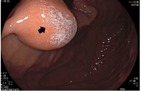
Figure 1 Upper gastrointestinal videoendoscopy showing a subepithelial tu mor below the cardia under retroflexed view (arrow)
An endoscopic ultrasound revealed a rounded heterogenous, with hypoechoic and hyperechoic areas land well-defined borders. The lesion reached the muscular layer without affecting it. There was no evidence of distant metastases (Fig. 2) in the multislice computed tomography scan.

Figure 2 Computed tomography scan of the thorax and abdomen in the coro nal view, showing a lesion within the gastroesophageal junction walls (arrow)
The patient was evaluated by an oncology board which decided the surgical resection through the combined transgastric approach (laparoscopic and endoscopic)
Surgical technique
The procedure started with the patient in the French position. After performing pneumoperitoneum using Veress needle, a 10-mm optical trocar (Opti-View Trocar®) was placed above the umbilicus. Then, two balloon-tipped trocars, 5 mm and 12 mm in diameter, were introduced through the midclavicular line below the right and left costal margins for the surgeon’s left-and right-hand working ports, respectively. A subxiphoid incision was made for introducing the Nathanson® liver retractor. Both balloon-tipped trocars were introduced into the stomach under direct endoscopic view through a small incision made with a monopolar scalpel in the anterior wall of the greater curvature at the level of the gastric body. The stomach was inflated with CO2 through the 12-mm intragastric port at a pressure of 8 mm Hg.
Under endoscopic guidance and retroflexed view, the lesion was gently tractioned with a 5-mm Endo Clinch grasper held with the surgeon’s left hand, and the resection was performed using one fire of mechanical stapler with a 60 mm load through the 12- mm port held in the surgeon’s right hand. The surgical specimen was placed in an endoscopic retrieval bag and an endoscopic grasper was then used to retrieve the specimen through the mouth. Finally, the small gastrotomies used for introducing the intragastric ports were closed with two loads of mechanical stapler (see online video).
The patient evolved without postoperative complications and was discharged 24 hours later tolerating solid food. The pathological examination of the surgical specimen reported complete surgical resection of a GIST with clear margins. One year after surgery the patient remains asymptomatic for dysphagia or gastroesophageal reflux.
The stomach is the most common location of GIST6. In 2008, Privette et al.7 described an easy classification based on submucosal gastric tumor location to manage the appropriate operative approach. GIST type I correspond to tumors located in the grater curvature and funds, type II are tumors of the prepyloric region and antrum and type III are tumors located in the lesser curvature or cardia, as the one described in this case report that was treated with the combined intragastric approach.
Several publications of case reports or case series have demonstrated that this approach is safe and valid8. A recent study by Barajas-Gamboa et al.9 demonstrated that laparo-endoscopic transgastric resection of gastric submucosal tumors was safe and efficient for GIST with optimal oncologic results.
The minimally invasive concept of these procedures has been emphasized by single incision laparoscopic surgery using single port devices for combined resections, with favorable outcomes10.
A possible limitation for this combined approach is the need for a group of professionals with both surgical and endoscopic skills. This association is essential for the treatment of many gastroesophageal conditions, which requires continuous training of surgeons endoscopists.
This technique has already been described and validated in the bibliography but is still innovative in our environment. Our experience is limited to few cases as the prevalence of these tumors in the GEJ is rare.
We have reported a case of a subcardial GIST resected using a combined laparoscopic and endoscopic approach. The procedure is safe, reproducible and relatively simple performed by trained professionals, representing a minimally invasive therapeutic option for patients with this condition.
Referencias bibliográficas /References
1. Mahajan NN, Sajan Jiv S N, Wong Kee Song LM, Blackmon SB. Lap aroendoscopicTransgastric Resection of Prepyloric Gastrointesti nal Stromal Tumor. Innovations (Phila). 2019; 14(1):66-8. [ Links ]
2. Goh BK, Chow PK, Chok AY, Chan WH, Chung YF, Ong HS, et al. Impact of the introduction of laparoscopic wedge resection as a surgical option for suspected small/medium-sized gastrointestinal stromal tumors of the stomach on perioperative and oncologic outcomes. J Gastrointest Surg. 2010; 34:1847-52. [ Links ]
3. Beltrán MA, Haito Y, Díaz R, Urbina O, Rodas C, De Balanzo A, Villa O. Resección mixta laparoscópica y endoscópica de un tumor del estroma gastrointestinal de la unión gastroesofágica. Rev Chil Cir. 2014; 66 (6): 586-9. [ Links ]
4. Tagaya N, Mikami H, Kogure H, Kubota K, Hoyosa Y, Nagai H. La paroscopic intragastric stapled resection of gastric submucosal tumors located near the esophagogastric junction. Surg Endosc. 2002; 16:177-9. [ Links ]
5. Ludwig K, Willhelm L, Scharlau U, Amtsberg G, Bernhardt J. Lapa roscopic- endoscopic rendezvous resection of gastric tumors. Surg Endosc. 2002; 16:1561-5. [ Links ]
6. Beltrán MA, Vicencio AO, Barra MM, Contreras MA, Wilson CS, Cruces KS. Resultados del tratamiento quirúrgico de los tumores del estroma gastrointestinal (GIST) en la IV Región de Chile. Rev Chil Cir. 2011; 63:290-6. [ Links ]
7. Privette A, McCahill L, Borrazzo E, Single RM, Zubarik R. Laparos copic approaches to resection of suspected gastric gastrointesti nal stromal tumors based on tumor location. Surg Endosc. 2008; 22:487-94. [ Links ]
8. Ismael H, Ragoza Y, Caccitolo J, Cox S. (2016). Optimal mana gement of GIST tumors located near the gastroesophageal junction: Case report and review of the literature. International Journal of Surgery Case Reports 2016; 25:91-6. doi:10.1016/j.ij scr.2016.06.006 [ Links ]
9. Barajas-Gamboa JS, Acosta G, Savides TJ, Sicklick JK, Fehmi SM, Coker AM, et al. Laparo-endoscopic transgastric resection of gas tric submucosal tumors. Surg Endosc 2015; 29(8):2149-57. [ Links ]
10. Na JU, Lee SI, Noh SM. The single incision laparoscopic Intragastric wedge resection of gastric submucosal tumors. J Gastric Cancer 2011;11:225-9. [ Links ]
Received: June 03, 2020; Accepted: September 11, 2020
 Este es un artículo publicado en acceso abierto bajo una licencia Creative Commons
