


 Inglês (pdf)
 Artigo em XML
 Referências do artigo
 Enviar este artigo por email
 Citado por SciELO
  Similares em
SciELO
Similares em
SciELO 
 Permalink
INTRODUCTION
Intracanal microbial reduction is the primary goal of root canal treatment, and is accomplished through irrigation, chemical debridement, and mechanical action of instruments 1 , allowing periradicular tissue healing. However, these steps can be difficult to complete due to the complexity of root canal anatomy 2 .
The internal canal configuration of mandibular canines has a high incidence of oval-shaped root canals 3 . Several rotary and reciprocating systems are used to promote complete cleaning of oval-shaped canals 4 , but leave unprepared areas after root canal instrumentaron 4-6 . Furthermore, anatomical complexities can also make it difficult to control infection during instrumentation, allowing accumulation of hard tissue debris, with microorganisms remaining in areas that instruments are unable to reach 4-6 . Remaining microorganisms might have the potential to perpetuate periapical inflammation and compromise the success of endodontic treatment 7 . Therefore, endodontic instruments with different kinematics and heat treatments have been developed to deal with root canals with complex anatomy, such as oval-shaped root canals 8 .
The WaveOne Gold system (Dentsply-Sirona, Ballaigues, Switzerland) is a reciprocating single-file made of a heat-treated gold metal alloy (M-wire) 9 , 10 . It has a triangular convex cross-sectional design with two cutting edges, resulting in one or two points of contact between the cutting edges and the dentin walls 9 , which can increase the flexibility and improve cyclic fatigue resistance when compared to conventional NiTi alloys 11 , 12 .
Mtwo is a well-known NiTi superelastic (SE) rotary system (VDW, Munich, Germany), with an “S”-shaped cross-sectional design, a positive rake angle with 2 cutting edges, and low radial contact to increase flexibility and improve performance during root canal prepararion 13 , 14 . Its shape enables dentin to be cut effectively and greater root canal residue removal 15 .
Therefore, the aim of this ex vivo study was to evaluate the shaping ability of single-file reciprocating (WaveOne Gold) and multifile rotary (Mtwo) systems on mandibular oval-shaped canine root canals, using microcomputed tomography (micro-CT). The null hypothesis tested was that there would be no difference between WaveOne Gold and Mtwo in (i) shaping ability or in (ii) apical transportaron and centering ability of mandibular oval-shaped canine root canals.
MATERIAL AND METHODS
This study was approved by the Iguaqu University Ethics Committee, Rio de Janeiro, Brazil (n.2.435.836).
Sample size calculation
A power calculation was performed based on data from a previous study 16 , with G*Power 3.1 software (Heinrich Heine University, Dusseldorf, Germany) using a power P = 95% and a = 5% as inputs into an independent samples test from the t tests family. The ideal sample size for each group was a minimum of 10 teeth. Five additional specimens per group were added to compensate for possible sample loss.
Specimen selection
Thirty mandibular canines with moderately curved mesial roots (10° to 20°) 17 were selected from a pool of 300 teeth from the Bank of Human Permanent Teeth of Iguaqu University. Teeth had been extracted for reasons unrelated to this study, Consent was secured prior to tooth donation. The teeth evaluated in this study were from patients of the metropolitan region of Rio de Janeiro city.
The remaining attached tissue was removed, and the teeth were stored in distilled water until the time they were to be used. All samples were scanned by micro-CT (SkyScan 1173, Bruker, Kontich, Belgium) operated at 50 kV and 160 mA, with a 1-mm-thick aluminum filter, 320-millisecond exposure time, 12.1 pm pixel size, 0.8 rotation step, and 360° rotation along the vertical axis. The files were then reconstructed into a three-dimensional dataset with the software NRecon v1.6.1.0 (Bruker micro-CT). Reconstruction parameters included a 50% beam hardening correction, ring artifact correction of 10, and fixed contrast limits (0 - 0.05) for all image stacks. The volume of interest extended from the cementoenamel junction to the apex of the root, resulting in the acquisition of 600 to 700 axial cross sections per sample.
Then, CTAn (v. 1.14.4, Bruker Micro-CT) and CTVol (v.2.2.1, Bruker Micro-CT) software were used to evaluate root canal morphological and 3D configuration. After that, the teeth were matched according to anatomical similarities of preoperative canal volume, canal surface area, and 3D configuration and randomly assigned to one of two groups (n-15) according to the instrument to be used during root canal preparation: Mtwo (VDW GmbH, Munich, German) or WaveOne Gold (Dentsply-Sirona, Ballaigues, Switzerland).
Root canal procedures
Endodontic accesses were performed with high-speed diamond (1014 HL; KG Sorensen, Sao Paulo, Brazil) and Endo Z burs (Dentsply-Sirona,
Ballaigues, Switzerland). A 10 K file (Dentsply-Sirona, Ballaigues, Switzerland) was used to determine apical patency, and the working length (WL) was considered 1 mm short of the apical foramen. A glide path was accomplished with a 15 K file (Dentsply Sirona) up to the WL.
The WaveOne Gold (Dentsply-Sirona) and Mtwo rotary (VDW GmbH) systems were activated with a VDW Silver motor (VDW GmbH, Munich, Germany), according to manufacturer’s instructions.
WaveOne Gold system
The WaveOne (WOG) primary (25/.07) was used in a reciprocating movement with an in-and-out pecking motion and an amplitude of 3 mm with light apical pressure until the WL was reached. After three movements, the instrument was removed from the canal and cleaned with a wet sterile gaze.
Mtwo system
The root canals were prepared using the sequence 10/.04, 15/.05, 20/.06, 25/.06 at 250 rpm with pecking motion, and small brushing movement with light apical pressure until the WL was reached.
An irrigation protocol was used for both groups. Root canal irrigation was performed with 2 mL of 2.5% sodium hypochlorite (NaOCl) with a 30-G Endo-Eze needle (Ultradent Products Inc; South Jordan, UT, USA) inserted until it was 2 mm from the WL. Final irrigation was performed with 2 mL of 2.5% NaOCl, 2 mL of17% EDTA (Mil Fórmulas, Rio de Janeiro, RJ, Brazil) for 1 min and 2 mL of 2.5% NaOCl. The root canals were dried with paper points, after which the teeth were scanned for a second time using the same parameters as mentioned above. A single experienced operator performed all procedures.
Micro-CT Evaluation
The teeth were submitted to a second micro-CT scan and reconstructed (NRecon) using the same parameters as described previously. The postoperative stacks of the root canals after preparation were registered with their respective preoperative stacks with an affine algorithm of the 3D Slicer software. The software ImageJ 1.50d (National Institutes of Health, Bethesda, MD, USA) was used to evaluate the initial and final volume (mm3), surface area (mm2), percentage of unprepared area, canal transportation and centering ability. The unprepared canal area was determined by calculating the number of static voxels (voxels present in the same position on the canal surface before and after instrumentation) divided by the total number of voxels present on the root canal surface 6 , according to the following formula:
Canal transportation and centering ratio were calculated at 3 cross-sectional levels (3-, 5-, and 7-mm distance from the apical foramen) using the following equations 18 :
Degree of canal transportation = (m 1 - m2) - (d 1 - d 2 ) Canal centering ratio = (m 1 - m2) - (d 1 - d 2 ) or (d 1 -
d 2 ) - (m 2 - m2),
where m1 is the shortest distance from the mesial of root canal to the mesial of the non-prepared canal, m2 is the shortest distance from the mesial of root canal to the mesial of the prepared canal, d1 is the shortest distance from the distal of root canal to the distal of the non-prepared canal, and d2 is the shortest distance from the distal of root canal to the distal of the prepared canal 18 .
Statistical analysis
The degree of homogeneity between the groups at baseline was confirmed through the analysis of initial volume and initial surface area of the root canals (p>0.05). Data distribution was verified for normality with the Shapiro-Wilk test. Due to the lack of normality, a Kruskal-Wallis test was used to compare intragroup transportation and centering ability parameters. The Mann-Whitney T test was used to compare canal transportation and centering ability between the same canal sections in different groups. The data were processed with Prism 7.0 (GraphPad Software, Inc., La Jolla, CA, USA) and expressed as the median, minimum and maximum values. The significance level was set at 5%.
RESULTS
The degree of homogeneity of the matched teeth regarding canal volume and surface area before root canal preparation was confirmed (p>0.05). No significant difference was found regarding the percentage of unprepared root canal areas between groups for the entire root canal or in the apical third (p>0.05). There was an increase in volume and surface area after root canal preparation compared to the initial sample in the groups tested. These results are described in Table 1 and Fig. 1.
No significant difference was observed in centering ability between the experimental groups (p>0.05). Canal transportation showed no statistically
significant differences in the intragroup comparison at the evaluated sections in either group (p>0.05). When each section was analyzed separately, WaveOne gold had less transportation than the MTwo file only at the 5 mm section (p<0.05). No statistical difference was found in centering ability at any of evaluated levels between groups (p>0.05). The total analyzed values are shown in Table 2.
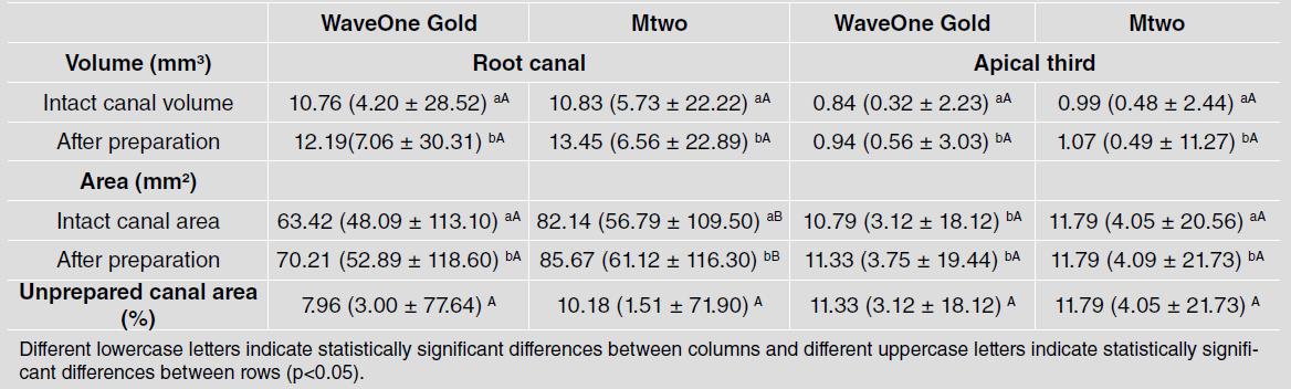
Table 1 Median, mínimum and máximum values of volume, surface area and percentage of unprepared canal area in root canal and apical third, after the different root canal preparations in WaveOne Gold and Mtwo Groups

Fig. 1 Representative 3D micro-CT images before (green) and after (red) root canal preparation of experimental groups: a) Wa-veOne Gold and b) MTwo. Representative transverse section of canals before (green) and after (red) root preparation at coronal (C), middle (M), and apical (A) thirds.
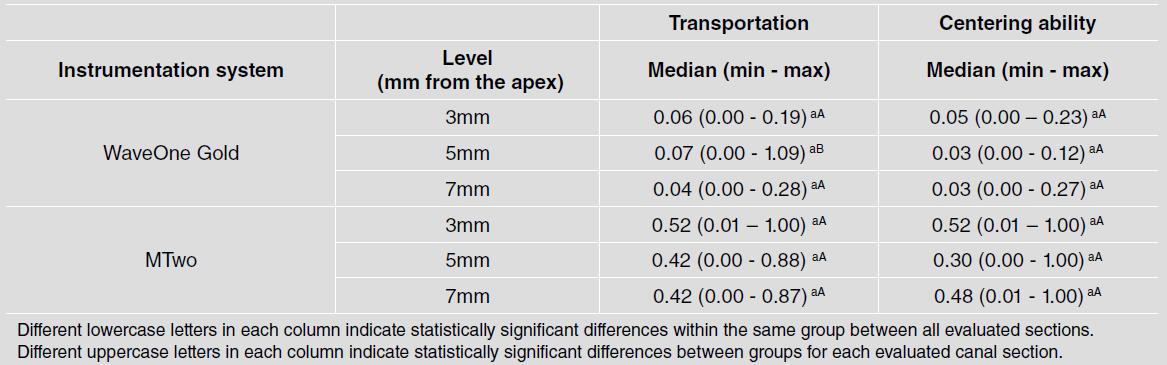
Table 2 Canal transportation and centering ability (mm) in the root canals sections after preparation for the two instrumentation systems
Different lowercase letters in each column indícate statistically significant differences within the same group between all evaluated sections. Different uppercase letters in each column indicate statistically significant differences between groups for each evaluated canal section.
DISCUSSION
The development of nickel-titanium (NiTi) rotary systems led to progress in root canal instrumentation 19 . However, failures may occur in oval and flattened canals because the instruments generally provide a rounded cross-section preparation, presenting a challenge to prepare all root canal walls. The instrumentation of these cases is more difficult due to the greater amount of dentin that must be removed to accomplish the ideal root canal shape 3 , 20 . The unprepared areas may harbor remnants of tissue and bacterial byproducts that could cause persistent infection and affect the success of endodontic treatment 21 .
Neither of the systems evaluated in this study was able to completely prepare the root canal, which agrees with previous studies 22-24 . Also, no significant difference was found for unprepared areas between WOG and Mtwo instruments, either in the entire root canal or in the apical third. Thus, the first hypothesis was accepted. These results can be attributed to the standardization of the apical third by the diameter of the instruments tested 25 , 26 .
NiTi instruments have led to significant progress in root canal preparation 27 . Centering ability was evaluated as described by Gambill et al. 18 , who defines centering ability as the ability of the endodontic instrument to remain on the central axis of the root canal. In the present study, no significant difference was observed in centering ability between experimental groups, which is in line with other studies 12 , 28 . Although our study showed similar shaping ability in general results, when each section was analyzed separately, WOG file had less transportation than the MTwo instrument at the 5 mm section from the apex, which partially rejects the second hypothesis. This result can be explained by the fact that WOG is a gold wire heat-treated instrument, while Mtwo is a NiTi SE instrument which does not have controlled memory. The thermally treated NiTi alloys present a higher percentage of martensitic phase, which is more flexible than conventional NiTi files, and may explain why there is less canal transportation of WOG at the 5 mm section from the apex 29 . The present study selected only long oval-shaped canals because they are considered a significant clinical challenge 30 . Moreover, the sample was selected through micro-CT analysis, which provides excellent pairing of teeth, reducing the anatomical bias related to heterogeneity of root canal morphology 4 . The micro-CT technique affords reliable results in the evaluation of data on 2D and 3D parameters of root canal preparation because it is a trustworthy, precise method for this kind of analysis 5 .
Based on our results, WaveOne Gold and Mtwo systems presented similar shaping ability and centering ability during oval-shaped root canal preparation. However, WOG presented less transportation than MTwo at the 5 mm section from the apex.

