


Servicios Personalizados
Revista

Articulo
 Inglés (pdf)
 Articulo en XML
 Referencias del artículo
 Enviar articulo por email
Indicadores
-
 Citado por SciELO
Links relacionados
-
 Similares en
SciELO
Similares en
SciELO
Compartir

 Permalink
Revista argentina de cardiología
versión On-line ISSN 1850-3748
Rev. argent. cardiol. vol.83 no.3 Ciudad Autónoma de Buenos Aires jun. 2015
BRIEF REPORT
Plasma VEGF level Changes With Exercise in Patients with Chronic Coronary artery Disease
Variaciones de los niveles plasmáticos de VEGF con el ejercicio en pacientes con enfermedad coronaria crónica
JUAN A. GAGLIARDIMTSAC, 1, 2, NEIVA MACIEL1, JOSÉ L. CASTELLANOMTSAC, 1, VERÓNICA MIKSZTOWICZ3, GABRIELA BERG3, RICARDO J. GELPIMTSAC, 4
Address for reprints: Dr. Juan A. Gagliardi - Pi y Margall 750 - 2° piso - (C1155ADP) Ciudad Autónoma de Buenos Aires, Argentina - Tel.-Fax 54-11 4121-0873 - e-mail: jgagliardi@fibertel.com.ar
MTSAC Full Member of the Argentine Society of Cardiology
1 Cardiology Division, Hospital General de Agudos “Dr. Cosme Argerich”
2 Researcher of the Ministry of Health, Government of the Autonomous City of Buenos Aires
3 Laboratory of Lipids and Lipoproteins of the Department of Clinical Biochemistry, School of Pharmacy and Biochemistry, Universidad de Buenos Aires
4 Institute of Cardiovascular Physiopathology, School of Medicine, Universidad de Buenos Aires
ABSTRACT
background: The aim of this study was to assess the effect of acute and programmed physical exercise on plasma VEGF levels in chronic stable coronary artery disease patients. Following baseline evaluation, 21 patients <75 years underwent an exercise stress myocardial perfusion scan (acute), and were then randomly assigned to perform programmed rehabilitation exercise or continue with their normal therapy. VEGF assessed by SPECT imaging significantly decreased after stress ergometry (from 49.59±6.06 to 31.83±5.62 pg/ml; p=0.021). At one month, it increased (70.90±14.44 pg/ml) though not significantly with respect to baseline values (p=0.1) and significantly with respect to immediate post exercise values (p<0.01). No significant changes were observed in VEGF at 3 months or when results were compared according to the presence of ischemia or programmed exercise.
Acute exercise induced a significant reduction in VEGF values, without differences between programmed exercise and the control group.
Key words: Coronary Artery Disease - Exercise - Vascular Endothelial Growth Factor A
RESUMEN
introducción: Con el objetivo de evaluar el efecto del ejercicio físico agudo y programado sobre los niveles plasmáticos de VEGF en pacientes coronarios crónicos estables, se estudiaron 21 pacientes < 75 años a los que luego de la evaluación basal se les realizó un estudio de perfusión miocárdica con esfuerzo (agudo) y posteriormente se asignaron en forma aleatoria a realizar ejercicios programados de rehabilitación o continuar con el tratamiento habitual.
Los valores de VEGF disminuyeron significativamente en el posesfuerzo de la ergometría de la SPECT (de 49,59±6,06 a 31,83±5,62 pg/ml; p = 0,021). Al mes, los valores aumentaron (70,90 ± 14,44 pg/ml) con tendencia no significativa respecto del valor basal (p=0,1) y significativamente respecto de los valores del posesfuerzo inmediato (p<0,01). No se observaron cambios significativos en los valores de VEGF a los 3 meses y tampoco al comparar los resultados según la presencia de isquemia o la realización de ejercicios programados.
El ejercicio agudo indujo una reducción significativa en los valores de VEGF, sin diferencias entre el ejercicio programado y el grupo control.
Palabras clave: Enfermedad coronaria - Ejercicio - Factor A de crecimiento endotelial vascular
Abbreviations
| DS | Difference or ischemia score | sPECt Single photon emission computed tomography |
| Es | Exercise score | VEGF Vascular endothelial growth factor |
| RS | Resting score |
Rev Argent Cardiol 2015;83:233-236. http://dx.doi.org/10.7775/rac.v83.i3.5490
Received: 11/17/2014 - Accepted: 12/01/2014
Furthermore, ischemia is a major stimulus for VEGF production and local amplification effects. (2, 3)
Although some authors have used intravenous or intracoronary VEGF injection to stimulate angiogenesis (4, 5) scheduled exercise and, perhaps, the pro-duction of small repeated ischemic stimuli could be useful mechanisms to increase endogenous VEGF and, therefore, promote the development of collateral circulation.
The aim of this study was to evaluate the effect of acute exercise, as that performed in exercise stress testing during a myocardial perfusion scan, on VEGF plasma levels in chronic stable coronary patients and then to compare the effect of scheduled exercise with a control group.
METHODS
The study included 21 patients<75 years, with stable coro-nary disease verified by angiography, previous infarction or positive perfusion studies of more than 6 months evolution, who had not participated in programmed exercise groups within the last 3 months.
After baseline evaluation, all patients underwent an exercise stress myocardial perfusion scan (ergometric test) with SPECT imaging. Subsequently, two randomized groups were formed: one with programmed exercise three times a week for 12 weeks and another that continued with standard medical treatment (control group).
Blood samples were obtained at baseline, during recovery, 30 minutes after exercise and at 1- and 3-month follow-up.
A commercial Quantikine (R&D, cat N° DVE00) kit, consisting of a sandwich enzyme immunoassay technique with an anti-human VEGF monoclonal antibody, was used for VEGF dosage. This technique allows VEGF dosage in concentrations ranging between 15.6 and 1000 pg/ml, with inter- and intra-assay coefficient of variation of about 5% and 8%, respectively.
The images obtained with 99m Tc-MIBI SPECT were analyzed in a 17-segment model, where each segment was assigned a score of 0=normal perfusion; 1=mild hypoperfu-sion; 2=moderate hypoperfusion; 3=severe hypoperfusion and 4=no perfusion. Resting score (RS), exercise score (ES) and difference or ischemia score (DS) were obtained. A DS≥2 was considered positive for ischemia.
Scheduled exercises were carried out in two weekly ses-sions in a rehabilitation center and a third session at the patients home.
statistical analysis
Categorical variables were expressed as frequency and percentage. Continuous variables were expressed as mean±standard deviation or standard error of the mean and median (interquartile range) according to their distribution.
The analysis of discrete variables was performed using the chi-square test and Fishers exact test as appropriate.
Continuous variables were compared using Student´s t test for two groups or the Kruskal-Wallis test according to their distribution. Intragroup continuous variables were compared using paired-t test. A p value<0.05 was consid-ered as statistically significant.
Ethical considerations
The protocol was approved by the institutional Research Ethics Committee and informed consent was obtained from
all participants.
RESULTS
Twenty-one patients were included in the study. Mean age was 62.5±7 years and 17 patients were men (80.9%). Risk factors were: hypertension in 17 patients (81.0%), diabetes in 2 (9.5%), dyslipidemia in 14 (66.7%) and smoking in 2 (9.5%). Eight patients had history of prior myocardial infarction (38.1%), 7 of angioplasty (33.3%) and 2 of previous surgery (9.5%).
Myocardial perfusion for ES and RS was 5 (0-11) and 0 (0-7.5), respectively. Nine patients had ischemia with a median DS of 4 (2-6), ES of 8 (5.5-19) and RS of 2 (0-16).
There was no difference in the result of exercise stress testing according to the presence of ischemia in the SPECT study. The maximum heart rate during ex-ercise was 120.3±16 bpm in patients with ischemia and 119.3±16 bpm in patients without ischemia (p=0.89).
Ten patients were assigned to programmed rehabilitation exercises and 11 to the control group.
VEGF results in acute exercise are summarized in Table 1. Gamma camera VEGF values after exercise stress decreased significantly (from 49.49±6.06 pg/ ml to 31.83±5.62 pg/ml, p=0.021). At 1 month, val-ues markedly increased compared to immediate post exercise stress results (70.90±14.44 pg/ml, p=0.007), with a non-significant trend relative to baseline values (p=0.10). At 3 months, a non-significant increase was observed with respect to baseline (p=0.12), with values similar to those measured at 1 month (Figure 1).
The analysis of myocardial perfusion data accord-ing to the presence of ischemia revealed results similar to those of the general population (see Table 1). There was no relationship between VEGF changes and the degree of ischemia observed in SPECT.
table 1. VEGF levels with acute exercise in the total study popu-lation (n=21) and in the subgroups according to the presence or absence of ischemia in perfusion studies
| Baseline | After exercise stress test | Month 1 | Month 3 | |
| mean | 49.59 | 31.83* | 31.83* | 67.48 |
| se | 6.06 | 5.62 | 5.62 | 13.31 |
| Without ¡schemia (n = 12) | ||||
| mean | 46.69 | 34.29* | 34.29* | 63.25 |
| se | 6.78 | 9.60 | 9.60 | 17.14 |
| Ischemia (n = 9) | ||||
| mean | 53.44 | 28.56 | 28.56 | 73.11 |
| se | 11.26 | 3.65 | 3.65 | 22.11 |
SE: Standard error of the mean.
* p = 0.021 vs. baseline; t p = 0.007 vs. after exercise stress test; $ p = 0.10 vs. baseline.
tt p = 0.05 vs.after exercise stress test; 44 p = 0.057 vs. baseline; § p = 0.07 vs. after exercise stress test
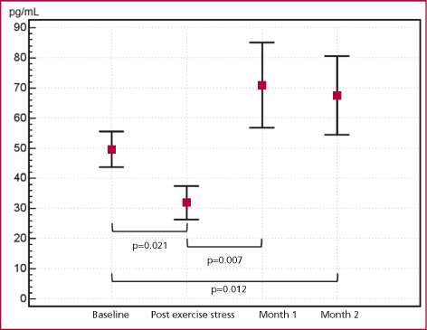
Fig. 1. Changes in VEGF levels (pg/ml) with acute exercise and at 1-month and 3-month follow-up.
significant differences at 1- and 3- month follow-up in patients who performed programmed cardiac rehabilitation exercises (Table 2).
There were no differences according to the presence of ischemia in the group of control patients or in that of programmed exercises.
DISCUSSION
VEGF serum values decreased significantly with acute exercise. The trend was similar when the analysis was performed according to the presence of ischemia.
Our results differ from those obtained in a study evaluating VEGF levels before and after exercise in coronary artery disease patients showing a slight non-significant decrease in its value. (6) In most of the studies evaluating a single maximal exercise session no significant changes in VEGF plasma levels were observed. (7-9)
The absence of variation in VEGF levels would be in line with data showing that exercise positively in-fluences angiogenesis which is, however, not directly determined by VEGF elevation but by its effect on VEGF rate. (6)
In acute myocardial infarction elevated VEGF serum levels were observed at 7 and 10 days of the acute event, (10) but other authors showed a significant re-duction immediately after reperfusion. (11) Thus, in the presence of an intense stimulus such as the acute ischemia of infarction, there would be an initial decline associated with reperfusion and perhaps a later elevation associated with the inflammatory and repair process, similar to that found with acute exercise test
table 2. VEGF levels at 1- and 3-month follow-up according to the performance of programmed physical exercises.
| Baseline Month 1 | Month 3 | |
| Control group (n = 11) | ||
| mean | 45.25 82.41 | 60.68 |
| se | 9.24 26.68 | 17.87 |
| Exercise group (n = 10) | ||
| mean | 54 <c-> 58.25 | 74.95 |
| se | 7.89 8.37 | 20.62 |
There were no signifcant differences. SE: Standard error of the mean. ing, especially in the presence of ischemia during ex-ercise stress.
An increase in VEGF values was detected at 1-month follow-up, without reaching statistical sig-nificance relative to baseline values.
This recovery was not very marked in patients who performed programmed exercises, as if routine exer-cise implied a permanent level of consumption, which was not exhibited in the control group.
The discrepancies observed with published data could result from the different times between exercise and blood withdrawal, and from the intensity and du-ration of both acute and chronic exercises.
limitations
This is a preliminary study with a small number of patients; therefore, further studies with a larger num-ber of patients should be undertaken to confirm these results.
CONCLUSIONS
Acute exercise produced a significant decrease of VEGF values, whereas chronic exercise did not determine significant changes with respect to the control group.
Conficts of interest
None declared.
(See author´s conflicts of interest forms in the web / Supple-mentary Material)
REFERENCES
1. Marino JC. Rehabilitación Cardiovascular. En: Bertolasi CA, editor. Cardiología 2000. Buenos Aires: Editorial Médica Panamericana; 1998. p. 1178-211.
2. Shintani S, Murohara T, Ikeda H, Ueno T, Honma T, Katoh A, et al. Mobilization of endothelial progenitor cells in patients with acute myocardial infarction. Circulation 2001;103:2776-9. http://doi. org/db75bp
3. Sandri M, Adams V, Gielen S, Linke A, Lenk K, Krankel N, et al. Effects of exercise and ischemia on mobilization and func-tional activation of blood-derived progenitor cells in patients with ischemic syndromes: results of 3 randomized studies. Circulation 2005;111:3391-9. http://doi.org/fswpxv
4. Hanif M, Patel A, Dunning J. Might gene therapy offer symptomatic relief for patients with no option angina? Interact Cardiovasc Thorac Surg 2005;4:627-32. http://doi.org/bq9ddf
5. Henry TD, Annex BH, McKendall GR, Azrin MA, Lopez JJ, Gior-dano FJ, et al. The VIVA trial: Vascular endothelial growth factor in Ischemia for Vascular Angiogenesis. Circulation 2003;107:1359-65. http://doi.org/b4cmx9
6. Danzig V, Mikova B, Kuchynka P, Benakova H, Zima T, Kittnar O, et al. Levels of circulating biomarkers at rest and after exercise in coronary artery disease patients. Physiol Res 2010;59:385-92.
7. Thijssen DH, Vos JB, Verseyden C, van Zonneveld AJ, Smits P, Sweep FC, et al. Haematopoietic stem cells and endothelial progenitor cells in healthy men: effect of aging and training. Aging Cell 2006;5:495-503. http://doi.org/fsgpx2
8. Van Craenenbroeck EM, Vrints CJ, Haine SE, Vermeulen K, Goo-vaerts I, Van Tendeloo V F, et al. A maximal exercise bout increases the number of circulating CD34+/KDR+ endothelial progenitor cells in healthy subjects. Relation with lipid profile. J Appl Physiol 2008;104:1006-13. http://doi.org/cbhdk3
9. Yang Z, Wang JM, Chen L, Luo C F, Tang AL, Tao J. Acute exer-cise-induced nitric oxide production contributes to upregulation of circulating endothelial progenitor cells in healthy subjects. J Hum Hypertens 2007;21:452-60.
10. Kranz A, Rau C, Kochs M, Waltenberger J. Elevation of vascular endothelial growth factor-A serum levels following acute myocardial infarction. Evidence for its origin and functional significance. J Mol Cell Cardiol 2000;32:65-72. http://doi.org/fdw6rm
11. Seko Y, Fukuda S, Nagai R. Serum levels of endostatin, vascular endothelial growth factor (VEGF) and hepatocyte growth factor (HGF) in patients with acute myocardial infarction undergoing early reperfusion therapy. Clin Sci (Lond) 2004;106:439-42. http://doi.org/ fgcnks

