


 Inglés (pdf)
 Articulo en XML
 Referencias del artículo
 Enviar articulo por email
 Citado por SciELO
  Similares en
SciELO
Similares en
SciELO 
 Permalink
INTRODUCTION
The insertion periodontium components that make up the topographic and functional unit are root cementum, periodontal ligament and alveolar bone. These structures evolve in an interrelated, coordinated manner over the life ofthe tooth, adapting continuously to changes that take place in the mouth. The periodontal ligament is located between the cementum and the alveolar bone. It is a fibrous connective tissue comprising a wide variety of cells: fibroblasts, osteoblasts, cementoblasts, osteoclasts, macrophages, mastocytes, undifferentiated ectomesenchymal cells and epithelial rests of Malassez (ERMs). Cell activity is mediated by several signaling factors that regulate the complex growth and regeneration machinery of periodontal structures 1 . After root formation, remnants of Hertwig’s sheath persist as ERMs throughout the life of the tooth 2 , 3 . Although their etiological role in the formation of odontogenic tumors and cysts is widely known, ERMs are currently a subject of interest as structures that take part in the histophysiology of the periodontal ligament. Histologically, ERMs are identified as small groups of epithelial cells within the periodontal ligament, in close contact with the cementum surface, becoming increasingly distant from the cementum surface towards the coronal third of the root 4 . Oblique sections of the periodontal ligament have shown ERMs to form part of mesh around the root 5 . ERMs have also been shown to have an irregular nucleus with dense heterochromatin and a small, scarcely distinguishable peripheral halo of cytoplasm 6 . ERM ultrastructure in sections of rat periodontal ligament is similar to that in humans and other animals 7 , 8 . A basal lamina separates the ERM cell islands from the connective tissue 9 , and there are tight junctions between them and hemidesmosomes, and tonofilaments within the cytoplasm. ERMs can produce different proteins and macromolecules, including cytokeratins 10 , 11 and neuropeptides 12 . The expression of CK 17 could be a marker used for identification 13 . Other studies 14 , 15 also report the expression of cell surface proteins such as epidermal growth factor receptors. It has been observed that proteins typical of the enamel matrix such as amelogenin and ameloblastin are expressed from samples of human periodontal ligament 16 , 17 . It has been suggested that these two proteins may act as growth factors and participate in adhesiveness, proliferation, migration and differentiation of periodontal ligament fibroblasts 18 , 19 . Moreover, it has been found that disruption of periodontal integrity induced early expression of APINA (another protein of the enamel matrix), forming part of a cascade of events possibly leading to the activation of the ERMs during periodontal healing and regeneration 20 .
Despite the ectodermal origin and epithelial nature of these cell groups, they can synthesize components that are frequently associated to cells ofmesenchymal origin, such as glycosaminoglycans, hyaluronic acid, dermatan sulfate and chondroitin sulfate 21 , as well as osteopontin (OPN), bone sialoprotein (BSP) and osteoprotegerin (OPG) 4 , 22 . They can also degrade collagen by synthesis of collagenases and proteinases 23 , 24 . It has therefore been suggested that they may contribute to periodontal regeneration by synthesizing a series of proteins related to the cementum and the bone tissues 25 . Current scientific evidence suggests that the possible role of ERMs in adult periodontal ligament is related to maintaining homeostasis of the periodontal space, thereby preventing anchylosis, inhibiting root resorption and contributing to cementum repair 26 , 27 .
In previous papers, our group has reported that in an experimental periodontitis model, ERMs exhibited cell hypertrophy when there was root resorption 28 . Other authors have evaluated ERM cell response to in vivo mechanical stimuli, observing cell proliferation and hypertrophy 29 . Regarding this point, our group studied ERMs in an experimental model of root resorption mediated by orthodontic forces in rats, finding no significant morphological change that would highlight their role in root resorption induced by orthodontic forces 30 . Other studies have described the expression of several proteins 27 , 31 that may contribute to the maintenance of cementogenesis and osteogenesis. Among these proteins, the expression of HSP 70 (Heat Shock Protein) by ERMs may provide protection against different forms of attack, including oxidizing agents, inflammation, hypoxia, hyperthermia and mechanical stimuli such as orthodontic forces 32 .
The aim of this study was to assess the behavior of periodontal tissues with relation to ERMs in healthy Wistar rats at different ages.
MATERIALS AND METHODS
We used 20 healthy male Wistar rats aged 1 month (n=7), 3 months (n=7) and 5 months (n=6). All animals had “ad libitum” access to balanced feed and water. They were housed in galvanized wire cages containing not more than 5 animals each, with temperature 21 °C to 24 °C; Humidity 52% to 56%; light/dark cycles: 12 hours/12 hours.
The experimental protocol was approved by the Ethics Committee of the School of Dentistry 012/2016 CICUAL-ODONTO-FOUBA of Buenos Aires, Argentina, and is in keeping with the National Institutes of Health Guidelines for the Care and Use of Laboratory Animals.
Histology and Histomorphometry
Following euthanasia at the relevant experimental times, lower jaws were extracted and fixed in 10% buffered formalin (pH 7.4). Samples were desiccated and decalcified in 10% EDTA solution, pH 7.0, for 25 days.
The section of each hemimandible corresponding to the three lower molars was embedded in paraffin to prepare mesiodistal histological sections ofthe lower first molar. Sections were stained with hematoxylin-eosin. The section with best orientation from each hemimandible, showing mesial and distal roots up to the apices, was selected.
The following parameters were evaluated at the furcation zone:
-
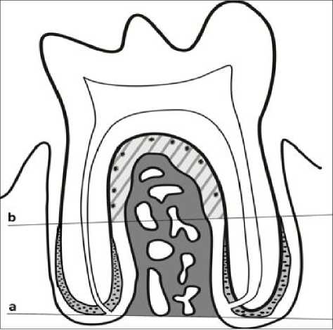
Number of ERMs/mm number of ERMs in the periodontal ligament in the perimeter limited by the beginning of cellular cementum, expressed with relation to root surface (Fig. 1).
Size/Area of ERMs (μm2) : Area of ERMs
-
Height of periodontal ligament (PL.h) (μm) : average of five equidistant linear measurements between the cementum surface of the furcation zone and the beginning of the bone tissue (Fig. 2).
-
Area of cementum in thefurcation zone : C.Ar (μm2): measurement of the area of cementum in the zone limited by the projection of two lines, on the mesial and distal root surfaces (Fig. 3).
Bone area B.Ar/T.Ar : Area of trabecular bone tissue in the zone selected for measuring ERMs, expressed as percentage of total area of interradicular bone (Fig. 1).
Number of ERMs was counted directly under 100x magnification with a Bausch &Lomb microscope. The rest of the histomorphometric measurements were taken using 100X digital microphotographs taken with a Nikon Eclipse photomicroscope and Image Pro-Plus Software.
Statistical analysis was performed with post-hoc Bonferroni and ANOVA, considering significance at p< 0.05.
RESULTS
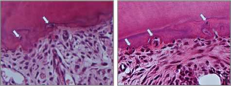
The results show that number of ERMs/mm declined significantly with age of animals, with major variability and high standard deviations in size of ERMs found in each group. This variability is reflected by the high standard deviations obtained for this parameter. In the 5-month-old group, ERMs were only observed in 3 of the 6 animals. Moreover, there was a significant increase in cementum area and bone area in the interradicular zone and a significant decrease in the height of the periodontal ligament in the 5-month-old group in relation to the other groups (Table 1 and Figs 4 and 5). Interestingly, we observed nuclei compatible with ERMs associated with and even embedded in the cementum matrix in the furcation zone (Fig. 6).
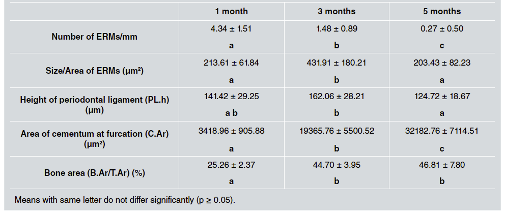
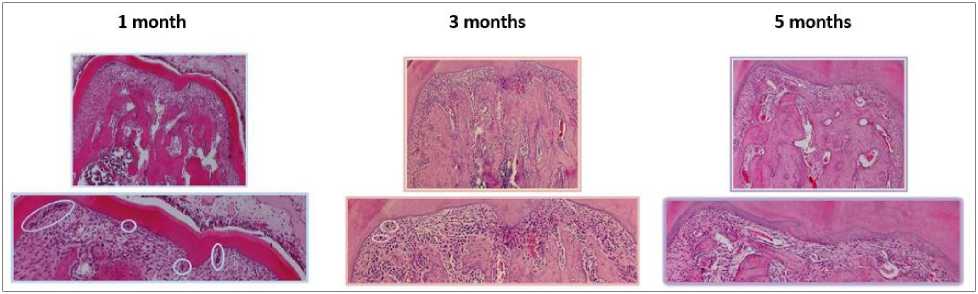
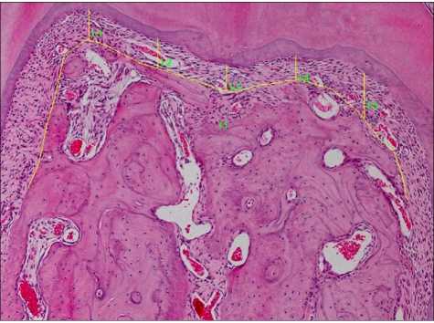
Fig. 4 Microphotographs of mesiodistal sections of lower molar of Wistar rats aged 1, 3 and 5 months, showing the decline in number of ERMs, the increase in cementum area at the level of the furcation and the reduction of the periodontal space as animal chronological age increases. H&E. 40 and 100X.
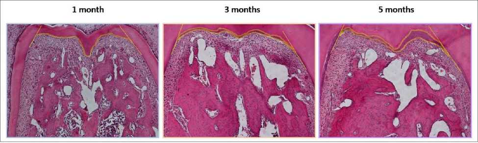
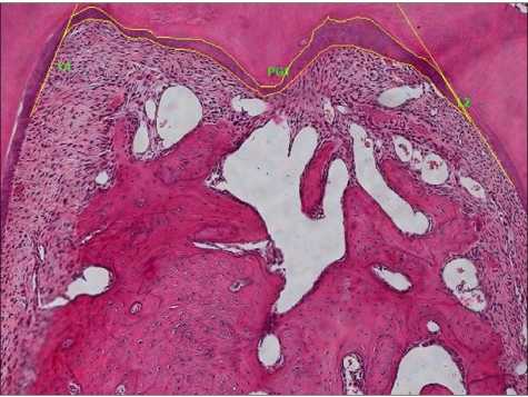
Fig 5 Microphotographs of mesiodistal sections of lower molar in Wistar rats aged 1, 3 and 5 months, showing delimitation established for evaluating the cementum area in the furcation zone in the 3 age groups. H&E.100X.
DISCUSSION
The results of this study show that the number of ERMs of lower molars in Wistar rats declines significantly as animal chronological age increases, and that there is major variability in size. The decline in number of ERMs was accompanied by a significant increase in interradicular cementum area at the level of the furcation and the interradicular bone area, entailing a reduction in height of the periodontal ligament when the animals were 5 months old.
The decline observed in number of ERMs with increase in age agrees with different papers published since 1950. Wentz et al.33 suggests that the decline in number of ERMs with age may be because they degenerate and then become calcified. Studies on human ERMs have also reported that they decline as age increases, and studies on older people report the onset of ERM calcification, which persist as cementum-like structures/cementicles34. A study by Gonçalves et al. 35 suggests that such decline may be partly due to cell apoptosis. In our study, the decline with age was accompanied by, and probably related to, an increase in the cementum area and interradicular bone volume and the consequent reduction in the height of the periodontal ligament.
Sims et al. 36 also reports a reduction in the thickness of the periodontal space as age increased in mice.
It is important to note that although our study did not find isolated partial calcifications in the periodontal ligament, it did find nuclei of cells compatible with possible ERMs embedded in the cementum associated to the interradicular space. The hypothesis that the nuclei observed in the radicular cementum matrix may have originated from nearby ERMs is also supported by the well-known fact that the topographic zone of the furcation and radicular cervical third is not characterized by presenting cellular cementum.
Hasegawa et al. 37 showed that during cementum repair, ERMs express osteopontin and ameloblastin, suggesting that they may be involved in cementum repair. More recent findings suggest that within the ERMs, there is a cell population with properties similar to those of undifferentiated mesenchymal cells, which, underthe right stimulus, can differentiate into cells capable of synthesizing mineralized matrices in a particular microenvironment 38 .
It has been more than twenty years since enamel matrix derivatives were introduced in clinical periodontics for use in regenerative therapies. Nevertheless, further research is still needed for better characterization of their effects on the behavior of different tissues and on the different cells 39 . In this context, it is worth noting that ERMs can express proteins typical of the enamel matrix, such as amelogenin and ameloblastin16. These proteins may have a strong effect on cell activity of the insertion periodontum 18 , 19 . Considering that ERMs may be a natural source of these proteins within the periodontal ligament itself, it is essential to continue learning about the behavior of ERMs in different contexts and under different stimuli.
The current study also found an increase in interradicular bone volume as chronological age increases, in agreement with other studies conducted by our group 40 . This finding poses a question that is yet unanswered, regarding whether there is any association between the increase in bone volume found and the behavior and/or presence of ERMs in the periodontal ligament. In further studies we will endeavor to ascertain whether there are mechanisms that operate in said association and how they work.
CONCLUSIONS
Further studies are needed to learn more about the role of ERMs and their association with the cementum, periodontal ligament and bone tissue, both in health and in situations requiring periodontal repair and/or regeneration mechanisms. In addition to providing insight regarding the biology of ERMs, this study contributes to a better characterization of an animal model that is extensively used in research on different aspects of oral pathology.

